

 PDF
PDF ePub
ePub Citation
 Print
Print


Abstract
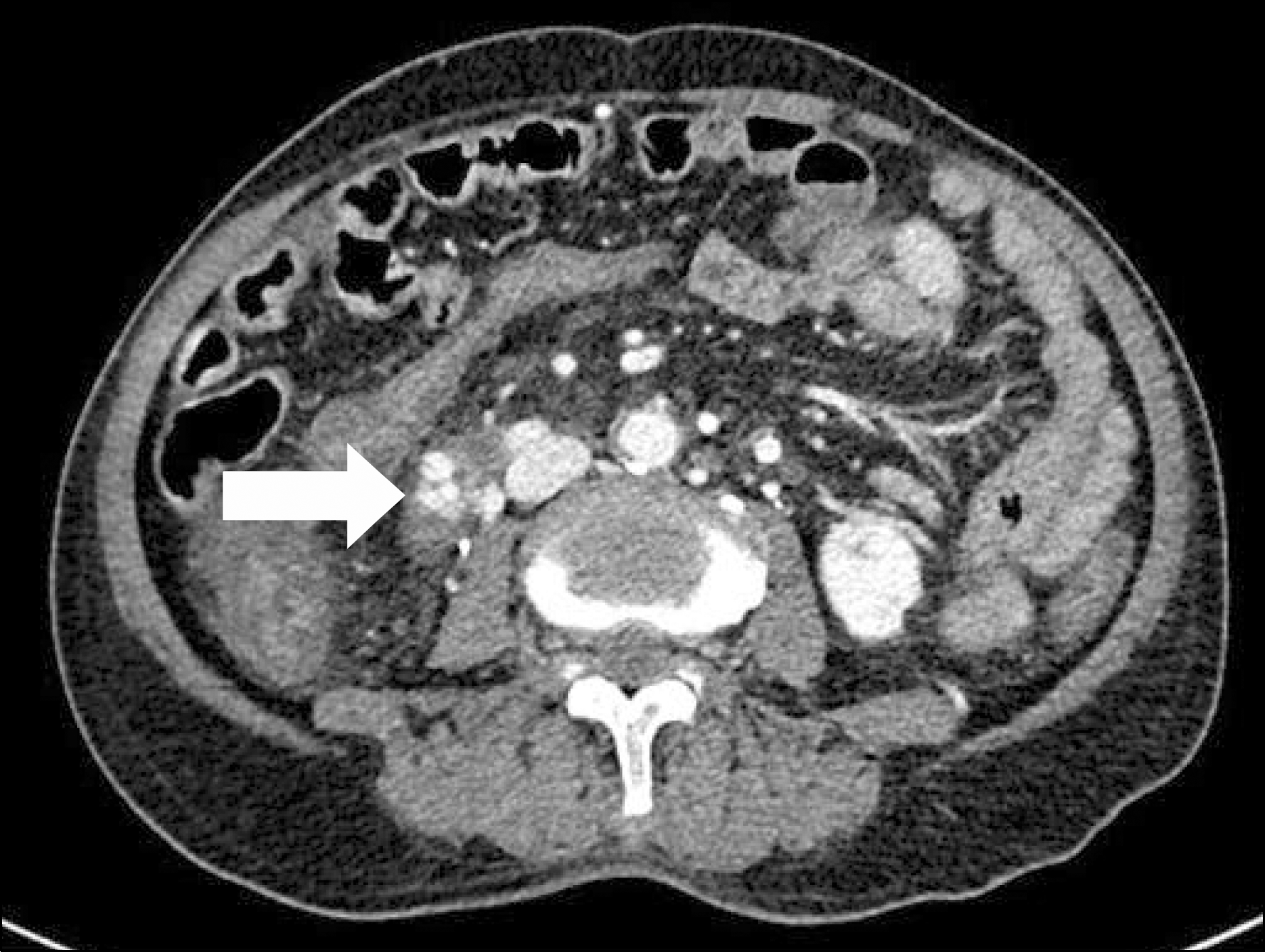
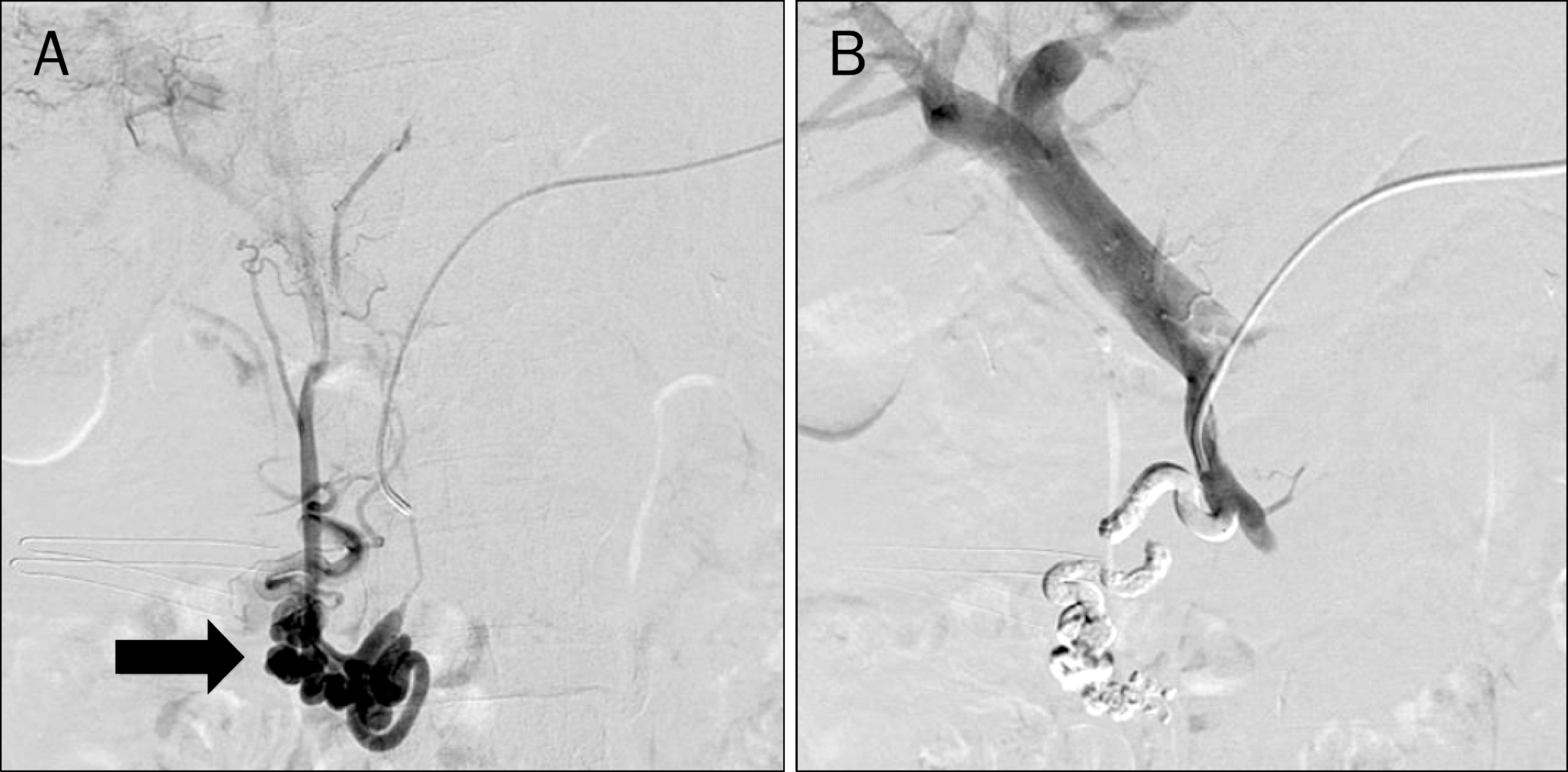
Variceal bleeding occurs primarily in the esophagus or stomach in patients with liver cirrhosis, but can also occur rarely in the duodenum. Duodenal variceal bleeding has a high mortality and poor prognosis due to heavy blood flow originating from the portal vein (PV) and the technical difficulty of hemostatic procedures. Treatments including endoscopic sclerotherapy, endoscopic ligations, endoscopic clipping and transjugular intrahepatic portosystemic shunt have been tried, with only moderate and variable success. A percutaneous transsplenic approach offers another way of accessing the PV. Here we report a case of successfully treated duodenal variceal bleeding by percutaneous transsplenic embolization.
References
1. Norton ID, Andrews JC, Kamath PS. Management of ectopic varices. Hepatology. 1998; 28:1154–1158.

2. Perera MT, Shimoda M, Kato M, et al. Life-threatening bleeding from duodenal varices due to pancreatic arteriovenous malformation: role of emergency pancreatoduodenectomy. Hepatogastroenterology. 2008; 55:1553–1556.
3. Khouqeer F, Morrow C, Jordan P. Duodenal varices as a cause of massive upper gastrointestinal bleeding. Surgery. 1987; 102:548–552.
4. Helmy A, Al Kahtani K, Al Fadda M. Updates in the pathogenesis, diagnosis and management of ectopic varices. Hepatol Int. 2008; 2:322–334.
5. Sato T, Akaike J, Toyota J, Karino Y, Ohmura T. Clinicopathological features and treatment of ectopic varices with portal hypertension. Int J Hepatol. 2011. DOI: doi: 10.4061/2011/960720.
6. Hotta M, Yoshida H, Mamada Y, et al. Successful management of duodenal varices by balloon-occluded retrograde transvenous obliteration. J Nippon Med Sch. 2008; 75:36–40.
7. Vidal V, Joly L, Perreault P, Bouchard L, Lafortune M, Pomier- Layrargues G. Usefulness of transjugular intrahepatic portosystemic shunt in the management of bleeding ectopic varices in cirrhotic patients. Cardiovasc Intervent Radiol. 2006; 29:216–219.
8. Luo J, Yan Z, Wang J, Liu Q, Qu X. Endovascular treatment for non-acute symptomatic portal venous thrombosis through intrahepatic portosystemic shunt approach. J Vasc Interv Radiol. 2011; 22:61–69.
9. Silva MA, Hegab B, Hyde C, Guo B, Buckels JA, Mirza DF. Needle track seeding following biopsy of liver lesions in the diagnosis of hepatocellular cancer: a systematic review and metaanalysis. Gut. 2008; 57:1592–1596.
10. Tung WC, Huang YJ, Leung SW, et al. Incidence of needle tract seeding and responses of soft tissue metastasis by hepatocellular carcinoma postradiotherapy. Liver Int. 2007; 27:192–200.
11. Brazzini A, Hunter DW, Darcy MD, et al. Safe splenoportography. Radiology. 1987; 162:607–609.
12. Probst P, Rysavy JA, Amplatz K. Improved safety of splenoportography by plugging of the needle tract. AJR Am J Roentgenol. 1978; 131:445–449.
13. Tuite DJ, Rehman J, Davies MH, Patel JV, Nicholson AA, Kessel DO. Percutaneous transsplenic access in the management of bleeding varices from chronic portal vein thrombosis. J Vasc Interv Radiol. 2007; 18:1571–1575.
14. Chu HH, Kim HC, Jae HJ, et al. Percutaneous transsplenic access to the portal vein for management of vascular complication in patients with chronic liver disease. Cardiovasc Intervent Radiol. 2012; 35:1388–1395.
15. Lee JY, Song SY, Kim J, et al. Percutaneous transsplenic embolization of jejunal varices in a patient with liver cirrhosis: a case report. Abdom Imaging. 2013; 38:52–55.
Fig. 1.
(A) Upper gastroduodenoscopy shows a varix in the second portion of duodenum, with red spot. (B) Endo-scopic photograph shows the successfully band-ligated duodenal varix.
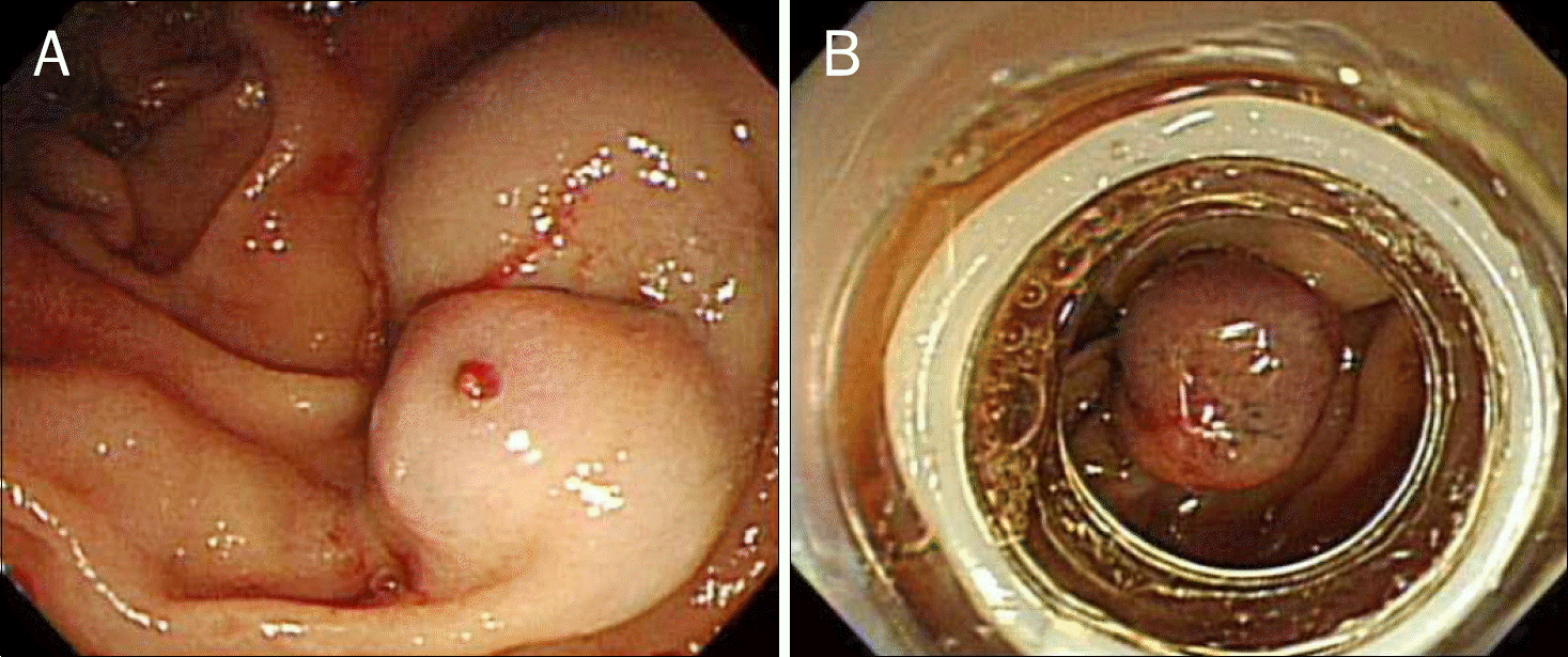
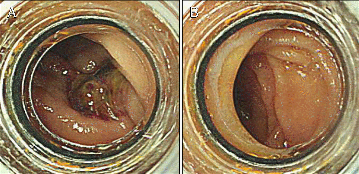
Fig. 2.
(A) On later endoscopy, post ligation ulceration was observed without bleeding sign. (B) There was no blood clot or trace of bleeding in other part of duodenum.
 XML Download