References
1. Yu JS, Kim KW, Lee HJ, Lee YJ, Yoon CS, Kim MJ. Urachal remnant diseases: spectrum of CT and US findings. Radiographics. 2001; 21:451–461.

2. Gearhart JP. Exstrophy, epispadias, and other bladder anomalies. Walsh PC, Retik AB, Vaughan ED, Wein AJ, Kavoussi LR, Novick AC, editors. Campbell's urology. 8th ed.Philadelphia: Saunders;2002. p. 2136–2196.
3. Berman SM, Tolia BM, Laor E, Reid RE, Schweizerhof SP, Freed SZ. Urachal remnants in adults. Urology. 1988; 31:17–21.

4. Iuchtman M, Rahav S, Zer M, Mogilner J, Siplovich L. Management of urachal anomalies in children and adults. Urology. 1993; 42:426–430.

5. Mesrobian HG, Zacharias A, Balcom AH, Cohen RD. Ten years of experience with isolated urachal anomalies in children. J Urol. 1997; 158:1316–1318.

Go to : 
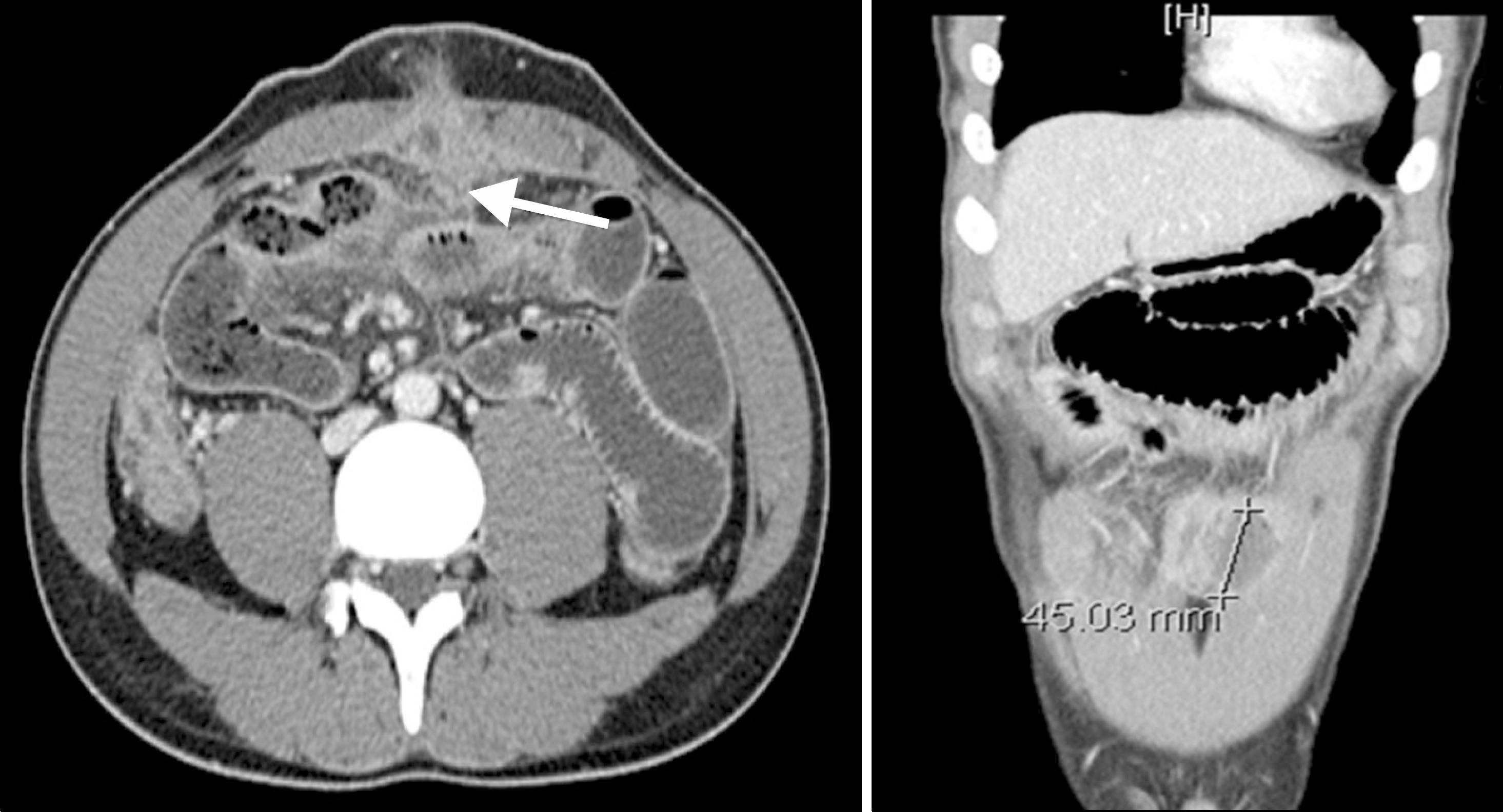
 | Fig. 1.Abdominopelvic CT findings. A 4.5 cm-sized abscess is seen beneath the mid-abdominal wall (arrow). Multisegmental strictures and diffuse small bowel dilatation, suggestive of small bowel obstruction due to stricture is noted. Multifocal bowel wall thickening involving the cecum, ascending colon, and small bowel loops are also present. There is abscess formation from urachal remnant resulting in secondary peritonitis. |


 PDF
PDF ePub
ePub Citation
 Print
Print


 XML Download