Abstract
Gastric cancer frequently disseminates to the liver, lung, and bone via hematogeneous, lymphatic, or peritoneal routes. However, gastric adenocarcinoma that metastasize to the colon and that shows typical linea platisca pattern on colonofiberscopy has rarely been reported. Recently, the authors experience a case of advanced gastric cancer with colonic metastases in a 55-year-old female patient. Multiple colonic lymphoid hyperplasias were detected on colonofiberscopy and biopsy revealed metastatic gastric cancer to the colonic wall. She was treated with mFOLFOX (5-FU, oxaliplatin, leucovorin) and has achieved stable disease status without disease progression. Herein, we report a rare case of signet ring-cell gastric cancer which metastasized to the colon in the form of multiple colonic lymphoid hyperplasias.
Go to : 
References
1. Batson OV. The function of the vertebral veins and their role in the spread of metastases. 1940. Clin Orthop Relat Res. 1995; (312):4–9.
2. Gao B, Xue X, Tai W, et al. Polypoid colonic metastases from gastric stump carcinoma: a case report. Oncol Lett. 2014; 8:1119–1122.

3. Lauren P. The two histological main types of gastric carcinoma: diffuse and so-called intestinal-type carcinoma. An attempt at a histoclinical classification. Acta Pathol Microbiol Scand. 1965; 64:31–49.
4. Eyres KS, Sainsbury JR. Large bowel obstruction due to metastatic breast cancer: an unusual presentation of recurrent disease. Br J Clin Pract. 1990; 44:333–334.
5. Sakai H, Egi H, Hinoi T, et al. Primary lung cancer presenting with metastasis to the colon: a case report. World J Surg Oncol. 2012; 10:127.

6. Tessier DJ, McConnell EJ, Young-Fadok T, Wolff BG. Melanoma metastatic to the colon: case series and review of the literature with outcome analysis. Dis Colon Rectum. 2003; 46:441–447.
7. Katon RM, Brendler SJ, Ireland K. Gastric linitis plastica with metastases to the colon: a mimic of Crohn's disease. J Clin Gastroenterol. 1989; 11:555–560.
8. Metayer P, Antonietti M, Oumrani M, Hemet J, Lemoine F, Basuyau J. Metastases of a gastric adenocarcinoma presenting as colonic polyposis. Report of a case. Dis Colon Rectum. 1991; 34:622–623.
9. Ogiwara H, Konno H, Kitayama Y, Kino I, Baba S. Metastases from gastric adenocarcinoma presenting as multiple colonic polyps: report of a case. Surg Today. 1994; 24:473–475.

10. Tiszlavicz L. Stomach cancer metastasizing into a solitary adenomatous colonic polyp. Orv Hetil. 1990; 131:1259–1261.
11. Tiszlavicz L. Metastasis of a stomach carcinoma in a solitary adenomatous cecal polyp. Zentralbl Allg Pathol. 1990; 136:277–282.
Go to : 
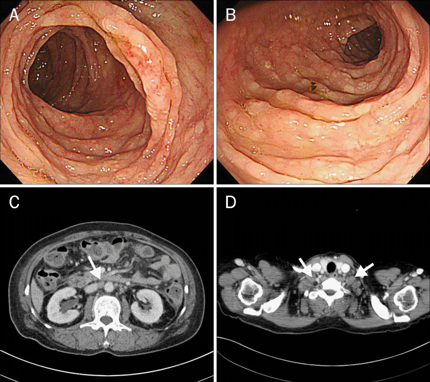
 | Fig. 1.Abdomen CT shows multiple enlarged perigastric lymph nodes (A), aortocaval, paraaortic, and portahe-patis lymph nodes (B). Chest CT shows metastatic supraclavicular lymph nodes (C) and lymphangitic metastasis at right apex (D). |
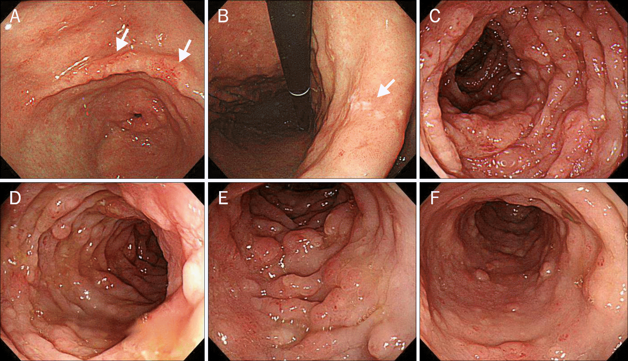
 | Fig. 2.(A, B) Upper endoscopy shows multiple shallow ulcers (arrows) with erythematous change on distal antrum and angle. (C-F) Colonoscopy shows multiple lymphoid hyperplasia from distal colon to rectum. |
 | Fig. 3.Biopsy specimens of the stomach and colon. H&E of stomach and colon shows infiltrating adenocarcinoma with signet ring cell feature (A, ×200; B, ×200). On immunohistochemical stain, colon specimen is positive for CK7 (C, ×200), and negative for CK20 (D, ×200) and CDX2 (E, ×200). Immunohistochemical stain of stomach is also positive for CK7 (F, ×200), and negative for CK20 (G, ×200) and CDX2 (H, ×200). |


 PDF
PDF ePub
ePub Citation
 Print
Print


 XML Download