

 PDF
PDF ePub
ePub Citation
 Print
Print


References
1. Caremani M, Vincenti A, Benci A, Sassoli S, Tacconi D. Ecographic epidemiology of non-parasitic hepatic cysts. J Clin Ultrasound. 1993; 21:115–118.

2. Choi BI, Lim JH, Han MC, et al. Biliary cystadenoma and cystadenocarcinoma: CT and sonographic findings. Radiology. 1989; 171:57–61.
3. Walt AJ. Cysts and benign tumors of the liver. Surg Clin North Am. 1977; 57:449–464.
4. Short WF, Nedwich A, Levy HA, Howard JM. Biliary cystadenoma. Report of a case and review of the literature. Arch Surg. 1971; 102:78–80.
5. Läuffer JM, Baer HU, Maurer CA, Stoupis C, Zimmerman A, Büchler MW. Biliary cystadenocarcinoma of the liver: the need for complete resection. Eur J Cancer. 1998; 34:1845–1851.
6. Sang X, Sun Y, Mao Y, et al. Hepatobiliary cystadenomas and cystadenocarcinomas: a report of 33 cases. Liver Int. 2011; 31:1337–1344.
7. Ishak KG, Willis GW, Cummins SD, Bullock AA. Biliary cystadenoma and cystadenocarcinoma: report of 14 cases and review of the literature. Cancer. 1977; 39:322–338.
8. Wang YJ, Lee SD, Lai KH, Wang SS, Lo KJ. Primary biliary cystic tumors of the liver. Am J Gastroenterol. 1993; 88:599–603.
9. Nakagawa M, Matsuda M, Masaji H, Goro W. Successful pre-operative diagnosis of biliary cystadenoma with mesenchymal stroma and its characteristic imaging features: report of two cases. Turk J Gastroenterol. 2011; 22:631–635.
10. Alobaidi M, Shirkhoda A. Malignant cystic and necrotic liver lesions: a pattern approach to discrimination. Curr Probl Diagn Radiol. 2004; 33:254–268.
11. Xu HX, Lu MD, Liu LN, et al. Imaging features of intrahepatic biliary cystadenoma and cystadenocarcinoma on B-mode and contrast-enhanced ultrasound. Ultraschall Med. 2012; 33:E241–E249.
12. Dixon E, Sutherland FR, Burak K, McKinnon G, May G. Cystadenoma of the liver without mesenchymal stroma in a female following hormonal therapy for acne. HPB (Oxford). 2001; 3:183–186.
13. Williams DM, Vitellas KM, Sheafor D. Biliary cystadenocarcinoma: seven year follow-up and the role of MRI and MRCP. Magn Reson Imaging. 2001; 19:1203–1208.
14. Suh JI, Kim JH, Lee DJ, et al. A case of biliary cystadenocarcinoma. Korean J Intern Med. 1997; 12:109–113.
15. Hai S, Hirohashi K, Uenishi T, et al. Surgical management of cystic hepatic neoplasms. J Gastroenterol. 2003; 38:759–764.
Go to : 
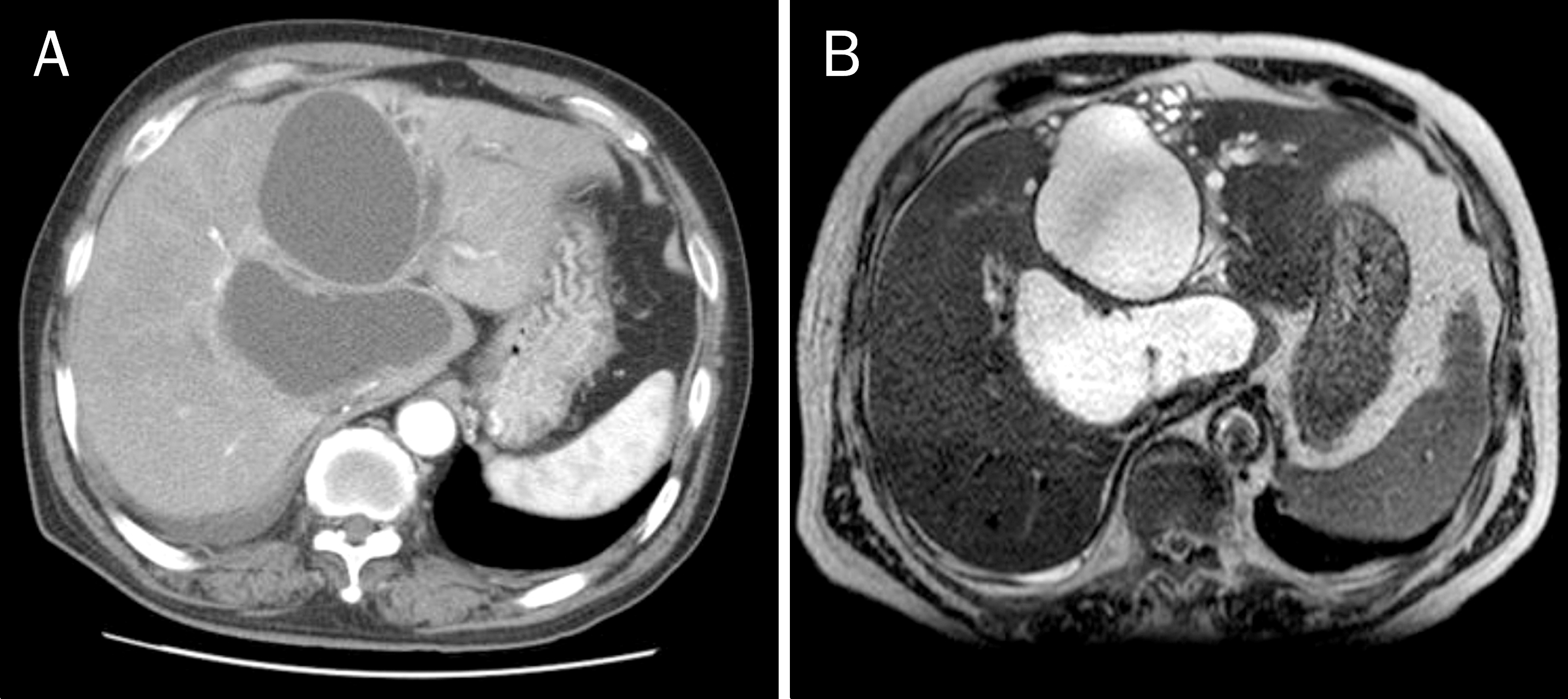 | Fig. 1.(A) Abdominal computed tomography scan shows large cystic lesions on arterial phase. (B) Magnetic reson-ance cholangiopancreatography scan also reveals large cystic lesions and adjacent small daughter cysts with high signal intensity on T2 weighted image without intramural nodules or septal wall thickening. |
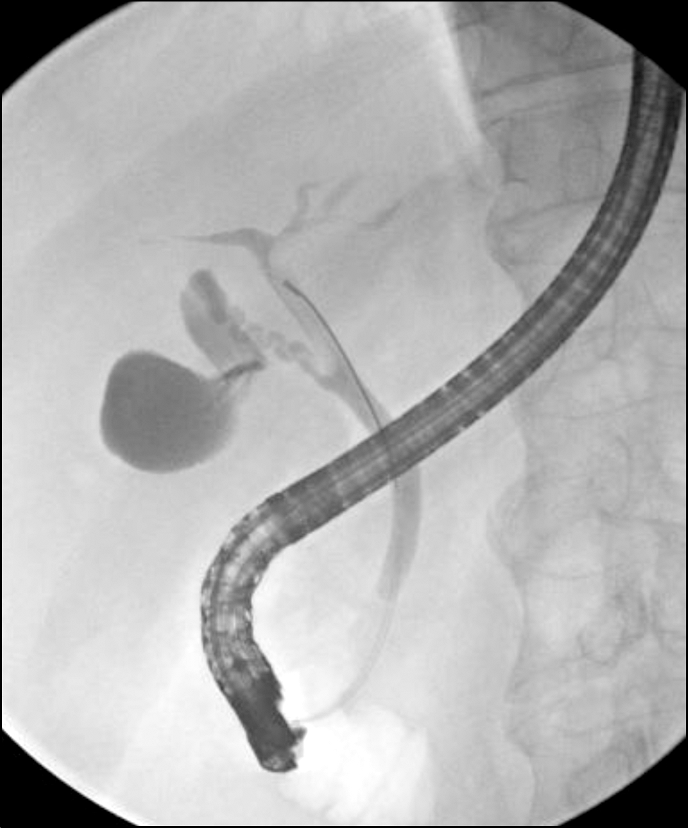 | Fig. 2.Endoscopic retrograde cholangiopancreatography shows a slight narrowing of common hepatic duct due to extrinsic compression and irregular stricture of intrahepatic duct without definite communication with cystic lesions. |
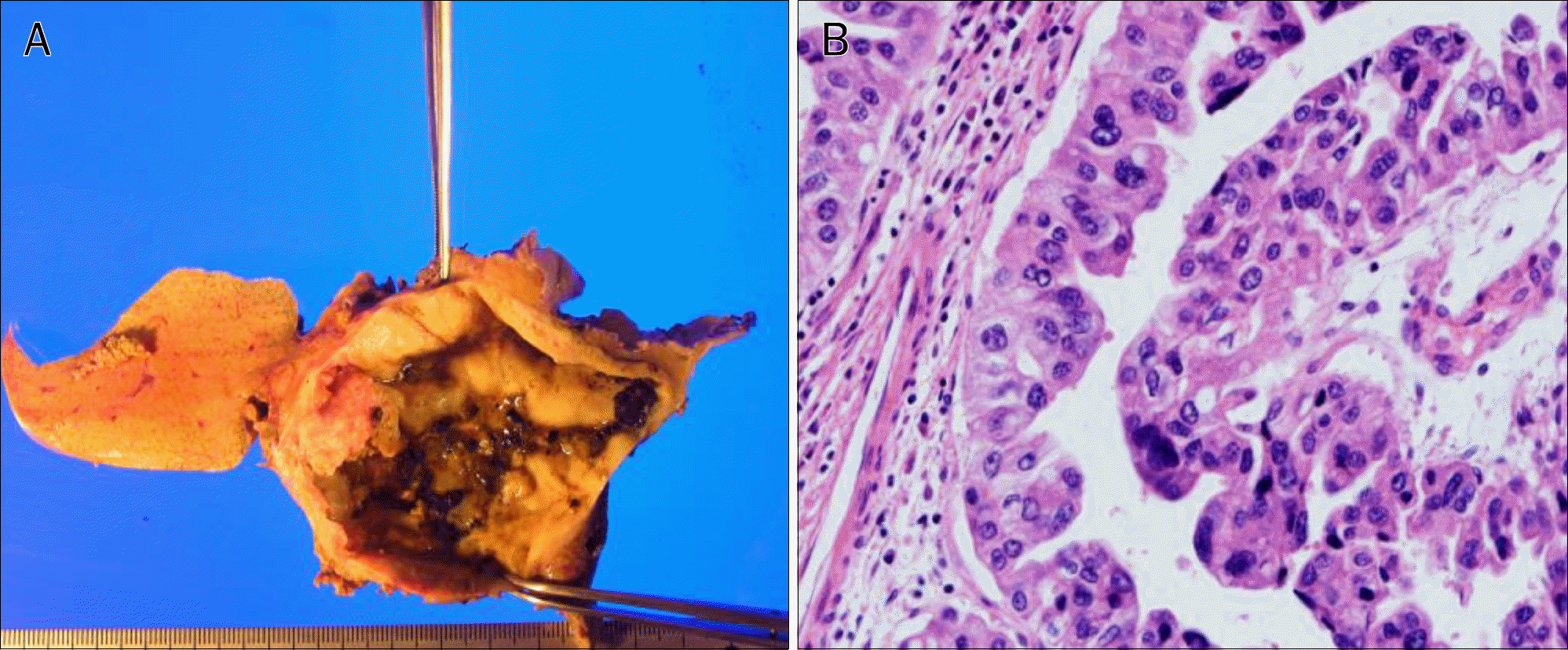 | Fig. 3.Macroscopic and microscopic findings of biliary cystadenocarcinoma. (A) Macroscopically, mural nodules, papillary infolding and septal thickening are observed within the largest cyst. (B) Microscopic finding shows cystadenocarcinoma with columnar epithelium and abundant cytoplasm containing mucin (H&E, ×400). |
 XML Download