

 PDF
PDF ePub
ePub Citation
 Print
Print


Abstract
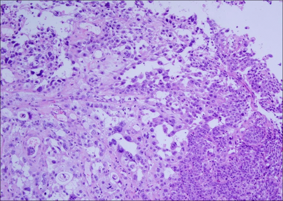
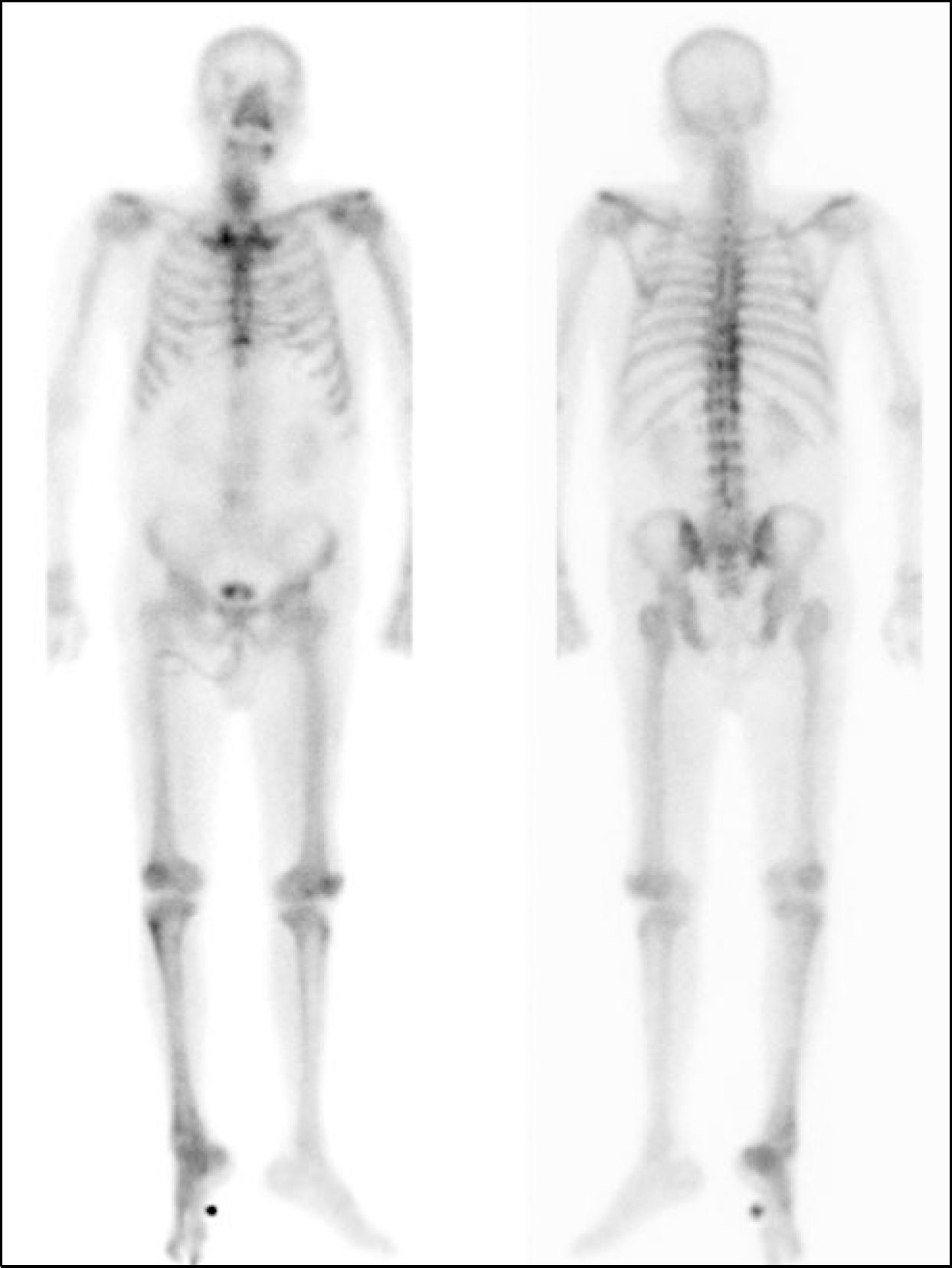
Paraneoplastic hypercalcemia without bone metastasis occurs rarely in esophageal cancer. A 75-year-old man was admitted for general weakness and lethargy. Laboratory data showed high serum calcium level (corrected calcium 14.6 mg/dL), low parathyroid hormone level (3.3 pg/mL) and high parathyroid hormone-related peptide level (3.5 pmol/L). Esophagogastroscopy showed a malignant tumor in the esophagus. Histology showed moderately differentiated squamous cell carcinoma. Bone scan showed no evidence of bone metastasis. Since the patient's calcium levels remained high and mental state did not show improvement despite intravenous fluid therapy, diuretics and intravenous bisphosphonate, hemodialysis was started. After hemodialysis treatment, the serum calcium level subsequently normalized and his mental status improved. Herein, we report a rare case of paraneoplastic hypercalcemia in a patient with esophageal cancer.
Go to : 
References
1. Vassilopoulou-Sellin R, Newman BM, Taylor SH, Guinee VF. Incidence of hypercalcemia in patients with malignancy referred to a comprehensive cancer center. Cancer. 1993; 71:1309–1312.

2. Lee JG, Han DS, Kim JH, et al. A case of adenocarcinoma of the transverse colon with humoral hypercalcemia of malignancy. Intest Res. 2012; 10:397–399.
3. Lim S, Han J, Park KH, et al. Two cases of humoral hypercalcemia of malignancy in metastatic cholangiocarcinoma. Cancer Res Treat. 2013; 45:145–149.
4. Kim JH, Kim BH, Lee DH, et al. A case of hepatocellular carcinoma presenting with four paraneoplastic syndromes such as hypercholesterolemia, hypoglycemia, hypercalcemia, and erythrocytosis. Korean J Gastroenterol. 2003; 41:316–320.
5. Yoon SY, Lee CR, Lee JH, Choi SJ, Son SP. A case of humoral hypercalcemia of malignancy associated with hepatoma: a case in which both PTHrP and 1,25 (OH) 2D were elevated. J Korean Soc Endocrinol. 1999; 14:197–202.
6. Stewart AF. Clinical practice. Hypercalcemia associated with cancer. N Engl J Med. 2005; 352:373–379.
7. Truong NU, deB Edwardes MD, Papavasiliou V, Goltzman D, Kremer R. Parathyroid hormone-related peptide and survival of patients with cancer and hypercalcemia. Am J Med. 2003; 115:115–121.
8. Reagan P, Pani A, Rosner MH. Approach to diagnosis and treatment of hypercalcemia in a patient with malignancy. Am J Kidney Dis. 2014; 63:141–147.
9. Clines GA. Mechanisms and treatment of hypercalcemia of malignancy. Curr Opin Endocrinol Diabetes Obes. 2011; 18:339–346.
10. Martin TJ, Moseley JM, Gillespie MT. Parathyroid hormone-related protein: biochemistry and molecular biology. Crit Rev Biochem Mol Biol. 1991; 26:377–395.
11. Li SH, Huang YC, Huang WT, et al. Is there a role of whole-body bone scan in patients with esophageal squamous cell carcinoma. BMC Cancer. 2012; 12:328.
12. Musat DL, Adhaduk M, Preminger MW, et al. Correlation of QT interval correction methods during atrial fibrillation and sinus rhythm. Am J Cardiol. 2013; 112:1379–1383.
13. Tachimori Y, Watanabe H, Kato H, et al. Hypercalcemia in patients with esophageal carcinoma. The pathophysiologic role of parathyroid hormone-related protein. Cancer. 1991; 68:2625–2629.
14. Watanabe HA, Matsushita H, Matsui H, et al. Esophageal carcinoma with high serum parathyroid hormone-related protein (PTHrP) level. J Gastroenterol. 1999; 34:510–515.
15. Nakajima N, Ueda M, Nagayama H, Yamazaki M, Katayama Y. Posterior reversible encephalopathy syndrome due to hypercalcemia associated with parathyroid hormone-related peptide: a case report and review of the literature. Intern Med. 2013; 52:2465–2468.
16. Nagashima R, Mabe K, Takahashi T. Esophageal small cell carcinoma with ectopic production of parathyroid hormone-related protein (PTHrp), secretin, and granulocyte colony-stimulating factor (G-CSF). Dig Dis Sci. 1999; 44:1312–1316.
Go to :
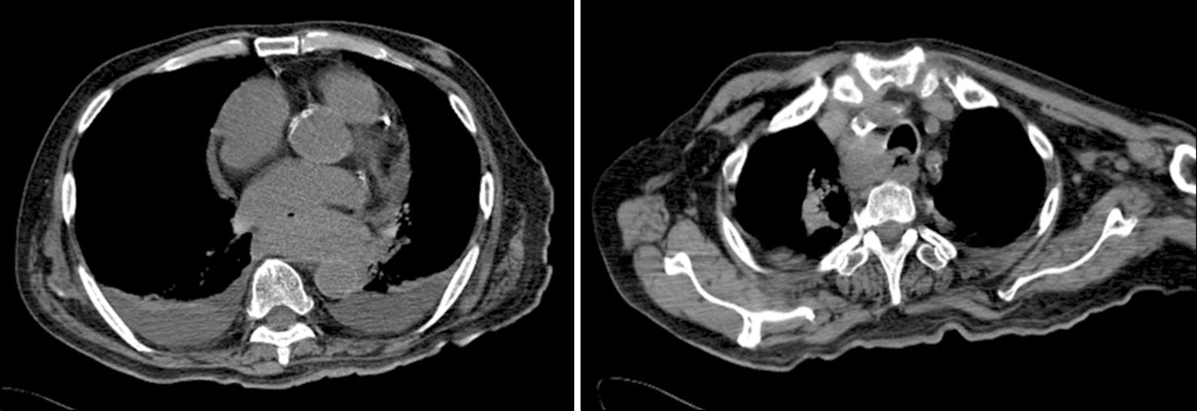 | Fig. 1.CT scan shows eccentric wall thickening of lower esophagus and paratracheal node enlargement. |
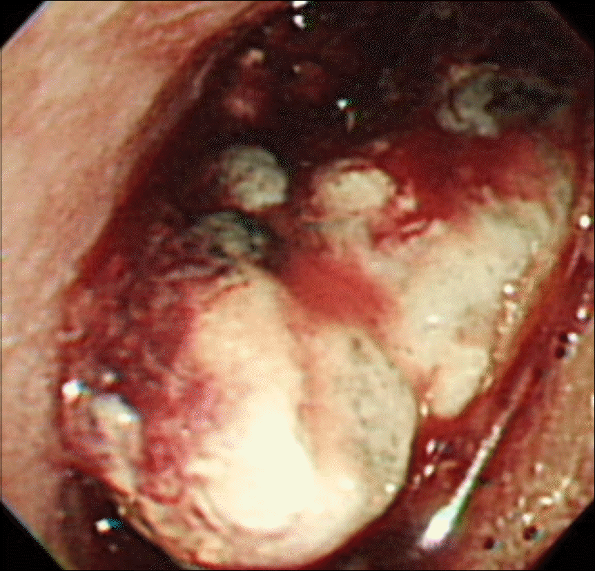 | Fig. 2.Esophagogastroscopy shows an ulcerative mass with bleeding and necrosis on the distal esophagus. |
 XML Download