References
1. Cooper GS, Xu F, Barnholtz Sloan JS, Schluchter MD, Koroukian SM. Prevalence and predictors of interval colorectal cancers in medicare beneficiaries. Cancer. 2012; 118:3044–3052.

2. Baxter NN, Sutradhar R, Forbes SS, Paszat LF, Saskin R, Rabeneck L. Analysis of administrative data finds endoscopist quality measures associated with postcolonoscopy colorectal cancer. Gastroenterology. 2011; 140:65–72.

3. Kim CJ, Jung YS, Park JH, et al. Prevalence, clinicopathologic characteristics, and predictors of interval colorectal cancers in Korean population. Intest Res. 2013; 11:178–183.

4. Bressler B, Paszat LF, Vinden C, Li C, He J, Rabeneck L. Colonoscopic miss rates for right-sided colon cancer: a pop-ulation-based analysis. Gastroenterology. 2004; 127:452–456.

5. Bressler B, Paszat LF, Chen Z, Rothwell DM, Vinden C, Rabeneck L. Rates of new or missed colorectal cancers after colonoscopy and their risk factors: a population-based analysis. Gastroenterology. 2007; 132:96–102.

6. van Rijn JC, Reitsma JB, Stoker J, Bossuyt PM, van Deventer SJ, Dekker E. Polyp miss rate determined by tandem colonoscopy: a systematic review. Am J Gastroenterol. 2006; 101:343–350.

7. Heresbach D, Barrioz T, Lapalus MG, et al. Miss rate for colorectal neoplastic polyps: a prospective multicenter study of back-to- back video colonoscopies. Endoscopy. 2008; 40:284–290.
8. Pickhardt PJ, Nugent PA, Mysliwiec PA, Choi JR, Schindler WR. Location of adenomas missed by optical colonoscopy. Ann Intern Med. 2004; 141:352–359.

9. Kahi CJ, Hewett DG, Norton DL, Eckert GJ, Rex DK. Prevalence and variable detection of proximal colon serrated polyps during screening colonoscopy. Clin Gastroenterol Hepatol. 2011; 9:42–46.

10. Pohl H, Srivastava A, Bensen SP, et al. Incomplete polyp re-section during colonoscopy-results of the complete adenoma resection (CARE) study. Gastroenterology. 2013; 144:74–80.e1.

11. Sawhney MS, Farrar WD, Gudiseva S, et al. Microsatellite in-stability in interval colon cancers. Gastroenterology. 2006; 131:1700–1705.

Go to : 
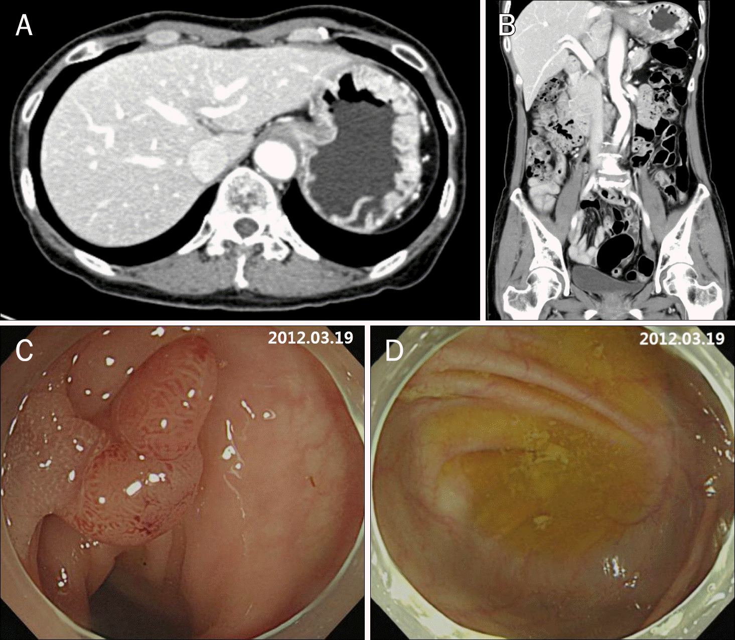
 | Fig. 1.Initial abdomen-pelvis CT and endoscopic findings. (A, B) No ab-normal findings are noted on abdo-men-pelvis CT (A, transverse view; B, coronal view). (C) About 10 mm sized, pedunculated polyp is observed on sigmoid colon and removed by endoscopic mucosal resection. (D) No ab-normal endoscopic findings are present on cecum. |


 PDF
PDF ePub
ePub Citation
 Print
Print


 XML Download