Abstract
Up-to-date imaging modalities such as three-dimensional dynamic contrast-enhanced CT (3D CT) and MRI may contribute to detection of hypervascular nodules in the liver. Nevertheless, distinguishing a malignancy such as hepatocellular carcinoma from benign hypervascular hyperplastic nodules (HHN) based on the radiological findings is sometimes difficult. Multiple incidental liver masses were detected via abdominal ultrasonography (US) in a 65-year-old male patient. He had no history of alcohol intake and no remarkable past medical history or relevant family history, and his physical examination results and laboratory findings were normal. 3D CT and MRI showed numerous enhanced nodules with hypervascularity during the arterial phase. After US guided liver biopsy, the pathological diagnosis was HHN. To date, several cases of HHN have been reported in patients with chronic alcoholic liver disease or cirrhosis. Herein, we report on a case of HHN in a patient with no history of alcoholic liver disease or cirrhosis.
Go to : 
References
1. Tani J, Miyoshi H, Sasaki M, et al. Multiple hypervascular FNH-like lesions in a patient with no history of alcohol abuse or chronic liver disease. Intern Med. 2013; 52:2225–2230.

2. Nakashima O, Kurogi M, Yamaguchi R, et al. Unique hypervascular nodules in alcoholic liver cirrhosis: identical to focal nodular hyperplasia-like nodules? J Hepatol. 2004; 41:992–998.

3. Park JE, Kim BS, Lee CH, Choi JH, Park YC, Park KK. A case of hypervascular hyperplastic nodules mimicking hepatocellular carcinoma in alcoholic liver cirrhosis. Korean J Hepatol. 2009; 15:193–200.

4. Park WK, Chang JC, Kim JW, et al. Hypervascular hyperplastic nodules appearing in chronic alcoholic liver disease: benign in-trahepatic nodules mimicking hepatocellular carcinoma. J Korean Radiol Soc. 2006; 54:113–119.

5. Moon JH, Ahn CM, Chung HS, Ahn SH, Park YN. A case of hypervascular hyperplastic nodules in a patient with alcoholic liver cirrhosis. Yonsei Med J. 2006; 47:881–886.

7. Namasivayam S, Salman K, Mittal PK, Martin D, Small WC. Hypervascular hepatic focal lesions: spectrum of imaging features. Curr Probl Diagn Radiol. 2007; 36:107–123.

8. International Working Party. Terminology of nodular hepatocellular lesions. Hepatology. 1995; 22:983–993.
9. Bosman FT. World Health Organization, International Agency for Research on Cancer. WHO classification of tumours of the digestive system. 4th ed.Lyon: IARC;2010. p. 196–261.
10. Kondo F. Benign nodular hepatocellular lesions caused by ab-normal hepatic circulation: etiological analysis and introduction of a new concept. J Gastroenterol Hepatol. 2001; 16:1319–1328.
11. Kim SR, Maekawa Y, Ninomiya T, et al. Multiple hypervascular liver nodules in a heavy drinker of alcohol. J Gastroenterol Hepatol. 2005; 20:795–799.

12. Maetani Y, Itoh K, Egawa H, et al. Benign hepatic nodules in Budd-Chiari syndrome: radiologic-pathologic correlation with emphasis on the central scar. Am J Roentgenol. 2002; 178:869–875.
Go to : 
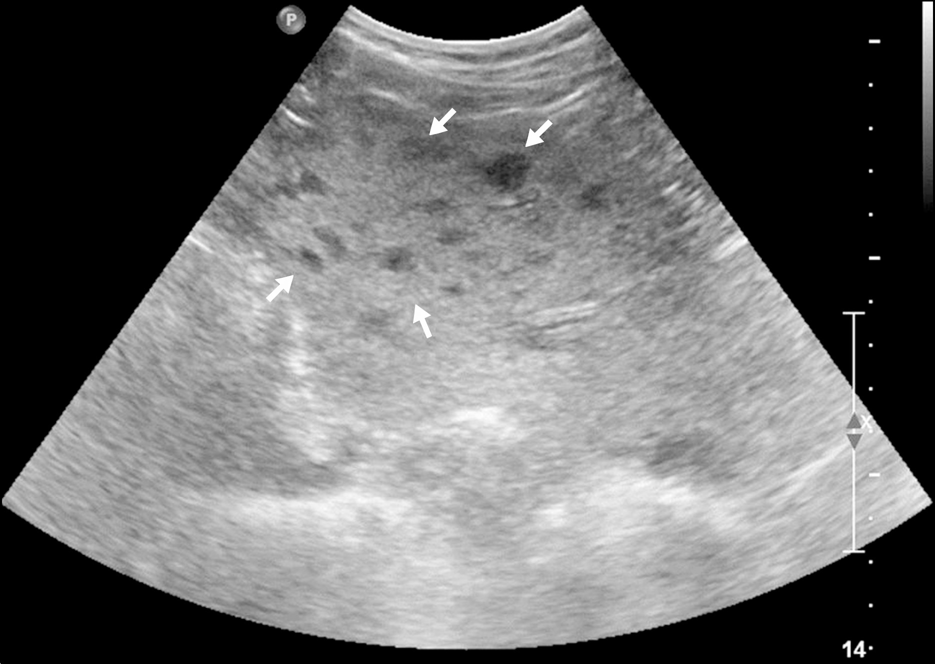
 | Fig. 1.Abdominal ultrasonography. Multiple-numerous hypoechoic nodules are seen in both lobes of the liver (arrows). |
 | Fig. 2.Abdominal three-dimensional dynamic contrast-enhanced CT images. (A) Pre-enhancement. Multiple nodules are highly enhanced in the arterial phase (B), with decreased enhancement in the portal phase (C), followed by iso-attenuation in the delayed phase (D). |
 | Fig. 3.Abdominal MRI. The hepatic nodules show iso-signal intensity on T1-weighted image (A) and T2-weighted image (B). The gadoxetate acid enhanced MR images obtained during the arterial (C) and portal phases (D) show the same findings shown on CT. |
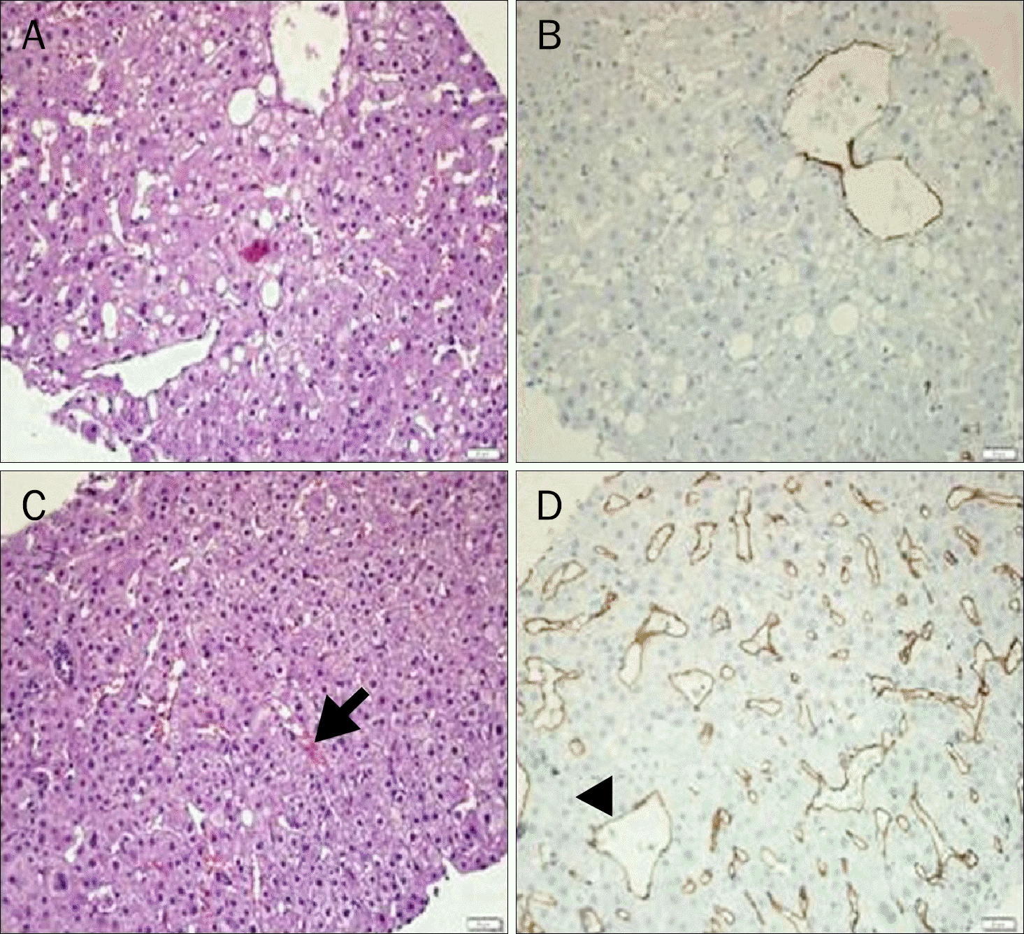
 | Fig. 4.Histopathology. (A) The non-nodular portion shows a normal parenchymal structure except for the mild fatty change (H&E stain, ×100).(B) The non-nodular portion shows little immunoreactivity for CD34 (CD34 stain, ×100). (C) The nodules have slightly increased cellularity without cellular atypia and mitotic figures, showing sinusoidal dilatation (arrow) with mild congestion (H&E stain, ×100). (D) The nodules show abundant sinusoidal capillarization and sinusoidal expression of CD34 (arrowhead) (CD34 stain, ×100). |


 PDF
PDF ePub
ePub Citation
 Print
Print


 XML Download