

 PDF
PDF ePub
ePub Citation
 Print
Print


Abstract
A 66-year-old male with dyspepsia and weight loss was referred to our hospital for evaluation. On laboratory examination, anti-saccharomyces cerevisiae (ASCA)-IgA was positive and iron deficiency anemia was present. PET/CT and abdominal CT scan images showed multiple small bowel segmental wall thickening and inflammation. Capsule endoscopy images showed multiple small bowel ulcerative lesions with exudates. Based on laboratory test results and imaging studies, the patient was diagnosed with Crohn's disease and treated with prednisolone and 5-aminosalicylic acid (5-ASA). However, the patient underwent second operation due to small bowel perforation within 2 month after initiation of treatment. Pathology report of the resected specimen was compatible to primary small bowel diffuse large B cell lymphoma and pertinent treatment was given to the patient after recovery. Herein, we describe a case of primary small bowel diffuse large B cell lymphoma that was mistaken for Crohn's disease.
References
1. Bernstein CN, Fried M, Krabshuis JH, et al. World Gastroenterology Organization Practice Guidelines for the diagnosis and management of IBD in 2010. Inflamm Bowel Dis. 2010; 16:112–124.

2. Saibeni S, Rondonotti E, Iozzelli A, et al. Imaging of the small bowel in Crohn's disease: a review of old and new techniques. World J Gastroenterol. 2007; 13:3279–3287.
3. Perlman SB, Hall BS, Reichelderfer M. PET/CT imaging of inflammatory bowel disease. Semin Nucl Med. 2013; 43:420–426.
4. Basso D, Zambon CF, Plebani M. Inflammatory bowel diseases: from pathogenesis to laboratory testing. Clin Chem Lab Med. 2014; 52:471–481.
5. Kim BG, Kim YS, Kim JS, Jung HC, Song IS. Diagnostic role of an-ti-Saccharomyces cerevisiae mannan antibodies combined with antineutrophil cytoplasmic antibodies in patients with inflammatory bowel disease. Dis Colon Rectum. 2002; 45:1062–1069.
6. Plevy S, Silverberg MS, Lockton S, et al. Combined serological, genetic, and inflammatory markers differentiate non-IBD, Crohn's disease, and ulcerative colitis patients. Inflamm Bowel Dis. 2013; 19:1139–1148.
7. Ibrahim EM, Ezzat AA, El-Weshi AN, et al. Primary intestinal diffuse large B-cell non-Hodgkin's lymphoma: clinical features, management, and prognosis of 66 patients. Ann Oncol. 2001; 12:53–58.
8. Kohli MD, Maglinte DD. CT enteroclysis in small bowel Crohn's disease. Eur J Radiol. 2009; 69:398–403.
9. Dave-Verma H, Moore S, Singh A, Martins N, Zawacki J. Computed tomographic enterography and enteroclysis: pearls and pitfalls. Curr Probl Diagn Radiol. 2008; 37:279–287.
10. Löffler M, Weckesser M, Franzius C, Schober O, Zimmer KP. High diagnostic value of 18F-FDG-PET in pediatric patients with chronic inflammatory bowel disease. Ann N Y Acad Sci. 2006; 1072:379–385.
11. Matysiak-Budnik T, Jamet P, Fabiani B, Nion-Larmurier I, Marjanovic Z, Ruskone-Fourmestraux A. Primary intestinal B-cell lymphoma: a prospective multicentre clinical study of 91 cases. Dig Liver Dis. 2013; 45:947–952.
12. Even-Sapir E, Lievshitz G, Perry C, Herishanu Y, Lerman H, Metser U. Fluorine-18 fluorodeoxyglucose PET/CT patterns of extranodal involvement in patients with non-Hodgkin lymphoma and Hodgkin's disease. Radiol Clin North Am. 2007; 45:697–709. vii.
13. Triester SL, Leighton JA, Leontiadis GI, et al. A meta-analysis of the yield of capsule endoscopy compared to other diagnostic modalities in patients with non-stricturing small bowel Crohn's disease. Am J Gastroenterol. 2006; 101:954–964.
14. Chermesh I, Eliakim R. Capsule endoscopy in Crohn's disease - Indications and reservations 2008. J Crohns Colitis. 2008; 2:107–113.
15. Shim KN, Kim YS, Kim KJ, et al. Korean Gut Image Study Group. Abdominal pain accompanied by weight loss may increase the diagnostic yield of capsule endoscopy: a Korean multicenter study. Scand J Gastroenterol. 2006; 41:983–988.
16. Dionisio PM, Gurudu SR, Leighton JA, et al. Capsule endoscopy has a significantly higher diagnostic yield in patients with suspected and established small-bowel Crohn's disease: a meta-analysis. Am J Gastroenterol. 2010; 105:1240–1248.
17. Hall CH Jr, Shamma M. Primary intestinal lymphoma complicating Crohn's disease. J Clin Gastroenterol. 2003; 36:332–336.
18. Vaidya R, Habermann TM, Donohue JH, et al. Bowel perforation in intestinal lymphoma: incidence and clinical features. Ann Oncol. 2013; 24:2439–2443.
Fig. 1.
(A) Conventional CT scan images show wall thickening and mucosal hyperenhancement on multiple segments of jejunum (arrows). (B) PET/CT scan images show increased uptake on multiple segments of jejunum.
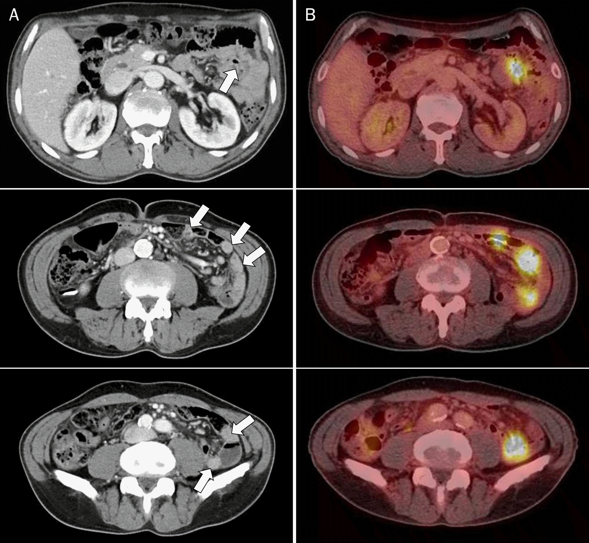
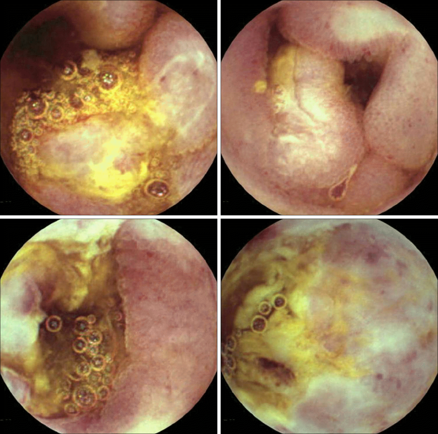
Fig. 3.
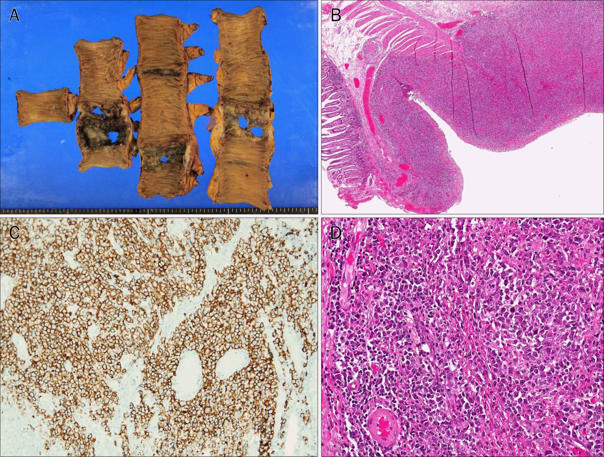
Gross and microscopic findings. (A) The resected jejunum shows multiple ulcers with perforations. (B) Microscopically, ill-defined ulcerative mass is seen throughout the jejunal wall (H&E, ×16). (C) Immunohistochemistry stain (CD20, ×100). CD20 is expressed in the tumor cells. (D) Large atypical lymphoid cell infiltration with non-cohesive pattern is observed on high-power field magnification (H&E, ×200).
 XML Download