

 PDF
PDF ePub
ePub Citation
 Print
Print


Abstract
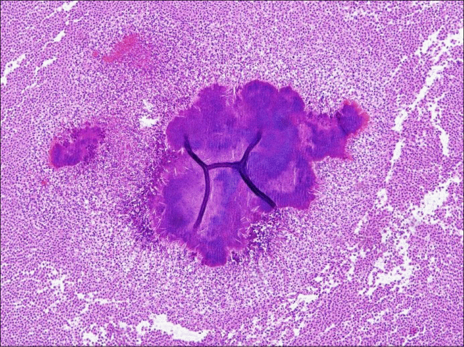
Actinomycosis is a chronic suppurative granulomatous infectious disease caused by actinomyces species that is characterized by formation of characteristic clumps called as sulfur granules. Abdominal actinomycosis is a rare disease and is often difficult to diagnose before operation. Abdominal actinomycosis infiltrating into the abdominal wall and adhering to the colon is even rarer. Most abdominal actinomycosis develops after operation, trauma or inflammatory bowel disease, and is also considered as an opportunistic infection in immunocompromised patient with underlying malignancy, diabetes mellitus, human im-munodefidiency virus infection, etc. Actinomycosis is diagnosed based on histologic demonstration of sulfur granules in surgically resected specimen or pus, and treatment consists of long-term penicillin based antibiotics therapy with or without surgical resection. Herein, we report an unusual case of abdominal wall actinomycosis which developed in a patient after acupuncture and presented as abdominal wall mass that was first mistaken for abdominal wall invasion of diverticulum perforation.
Go to : 
References
1. Choi PS, Kim SJ, Jeon HS, et al. A case of pelvic and abdominal actinomycosis associated with wearing an intrauterine device. Korean J Obstet Gynecol. 2001; 44:1357–1361.
2. Bae TS, Bae JD, Kim SO, Lee MS, Jung KH, Jung BW. Three cases of abdominal actinomycosis. J Korean Surg Soc. 2000; 59:414–419.
3. Ormsby AH, Bauer TW, Hall GS. Actinomycosis of the cholecystic duct: case report and review. Pathology. 1998; 30:65–67.

4. Cummins CS. Chemical composition and antigenic structure of cell walls of Corynebacterium, Mycobacterium, Nocardia, Actinomyces and Arthrobacter. J Gen Microbiol. 1962; 28:35–50.
5. Cope Z. Actinomycosis. London: Oxford University press;1938.
6. Putman HC Jr, Dockerty MB, Waugh JM. Abdominal actino-mycosis; an analysis of 122 cases. Surgery. 1950; 28:781–800.
7. Peabody JW Jr, Seabury JH. Actinomycosis and nocardiosis. A review of basic differences in therapy. Am J Med. 1960; 28:99–115.
8. Acquaro P, Tagliabue F, Confalonieri G, Faccioli P, Costa M. Abdominal wall actinomycosis simulating a malignant neoplasm: case report and review of the literature. World J Gastrointest Surg. 2010; 2:247–250.
9. Cintron JR, Del Pino A, Duarte B, Wood D. Abdominal actinomycosis. Dis Colon Rectum. 1996; 39:105–108.
10. Ha HK, Lee HJ, Kim H, et al. Abdominal actinomycosis: CT findings in 10 patients. AJR Am J Roentgenol. 1993; 161:791–794.
11. Goldwag S, Abbitt PL, Watts B. Case report: percutaneous drainage of periappendiceal actinomycosis. Clin Radiol. 1991; 44:422–424.
12. Chin SJ, Lee CK, Kim CW, Kim KH. Actinomycosis of liver and greater omentum: a case report. Korean J Pathol. 1979; 13:303–308.
13. Lambroza A, Tighe MK, DeCosse JJ, Dannenberg AJ. Disorders of the rectus abdominis muscle and sheath: a 22-year experience. Am J Gastroenterol. 1995; 90:1313–1317.
14. Kim JW, Jeong JB, Jung YJ, et al. Clinical features and therapeutic responses of abdominal actinomycosis. Intest Res. 2007; 5:177–183.
15. Nayak L, DiMaio M, Jeffrey RB. Extraglandular extension of paro-tid actinomycosis after sonographically guided fine-needle aspiration. J Ultrasound Med. 2013; 32:715–716.
Go to :
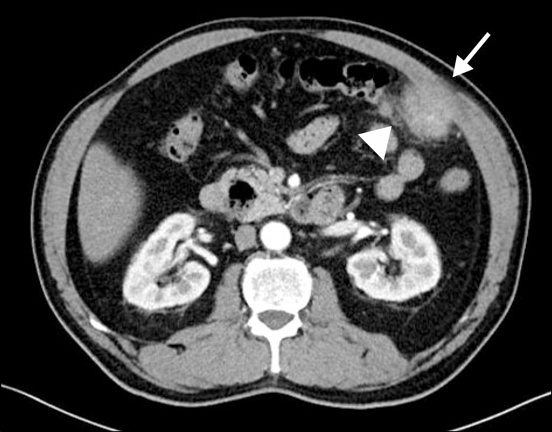 | Fig. 1.CT image shows an ill-defined, heterogeneous enhancing mass at the left upper quadrant of the abdomen that is focally infiltrating into the abdominal wall (arrow) and closely abutting adjacent transverse colon (arrowhead). |
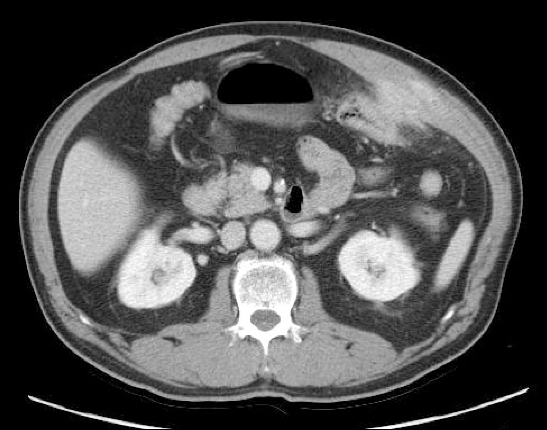 | Fig. 2.Follow-up computed tomography after 4 months shows increased size of enhancing mass with interval progression of infiltration into the abdominal wall and presumably to the transverse colon. Prominent peri-lesional infiltration is also demonstrated. |
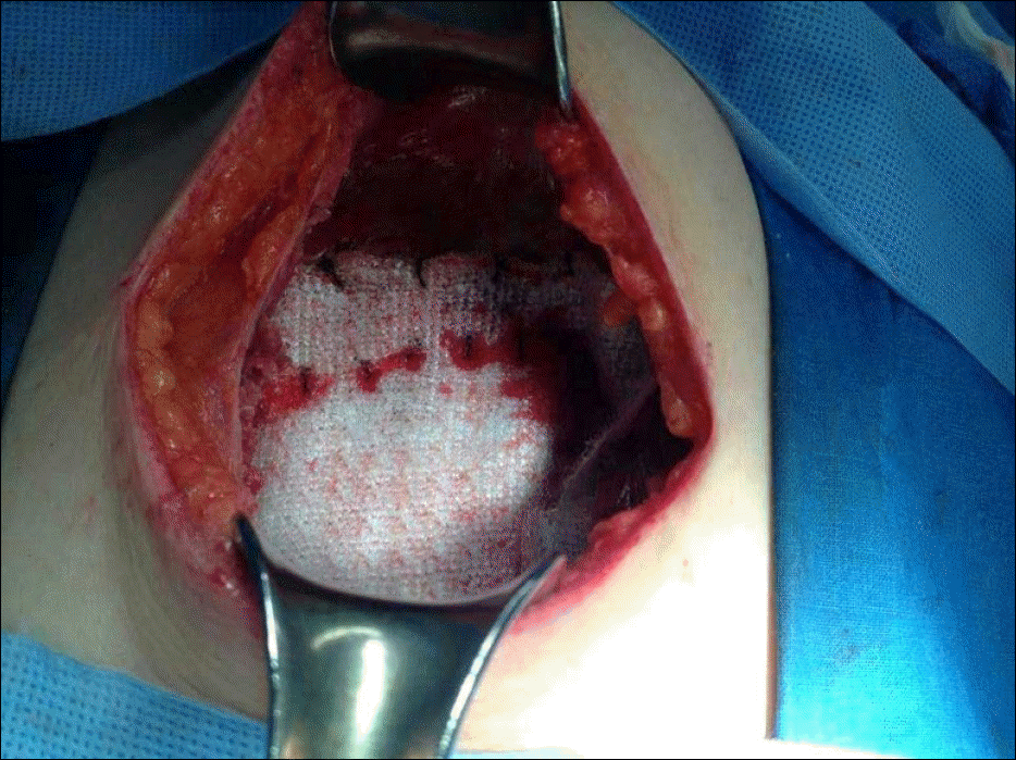
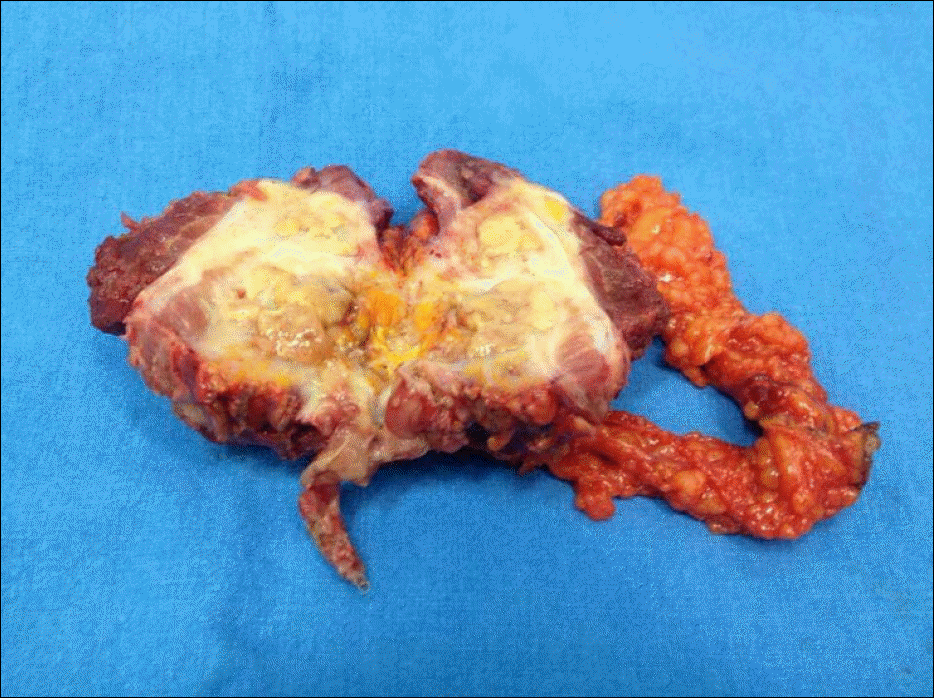 | Fig. 4.Gross finding of surgically resected specimen of abdominal wall actinomycosis conjoined with transverse colon which shows central yellowish necrosis. |

 XML Download