Abstract
Colonoscopy is a safe procedure performed routinely worldwide. Splenic rupture is a rare complication of colonoscopy with several reported cases since 1974. We report the first case of a complication in the Republic of Korea. The literature on this rare complication is also reviewed here, with focus on the analysis of risk, diagnosis, and treatment. A 77-year-old patient receiving oral aspirin underwent colonoscopy with polypectomy. After 24 hours, the patient experienced dizziness and hypotension. Colonoscopy was performed to exclude intestinal bleeding, which could be diagnosed with hemoperitoneum. A computed tomography scan showed copious abdominal free blood and a splenic rupture. An urgent splenectomy was performed, which was the recognized procedure of choice. Physicians should have greater awareness of the possibility of splenic rupture following colonoscopy in order to avoid delay of diagnosis and treatment for this life-threatening complication.
References
1. Macrae FA, Tan KG, Williams CB. Towards safer colonoscopy: a report on the complications of 5000 diagnostic or therapeutic colonoscopies. Gut. 1983; 24:376–383.

2. Wexner SD, Garbus JE, Singh JJ. SAGES Colonoscopy Study Outcomes Group. A prospective analysis of 13,580 colonoscopies. Reevaluation of credentialing guidelines. Surg Endosc. 2001; 15:251–261.
3. Garancini M, Maternini M, Romano F, Uggeri F, Dinelli M. Are there risk factors for splenic rupture during colonoscopy? Case report and literature review. J Gastroint Dig Syst. 2011; S2:001. doi:10.4172/2161-069X.S2-001.
4. Wherry DC, Zehner H Jr. Colonoscopy-fiberoptic endoscopic approach to the colon and polypectomy. Med Ann Dist Columbia. 1974; 43:189–192.
5. Moses RE, Leskowitz SC. Splenic rupture after colonoscopy. J Clin Gastroenterol. 1997; 24:257–258.

6. Jentschura D, Raute M, Winter J, Henkel T, Kraus M, Manegold BC. Complications in endoscopy of the lower gastrointestinal tract. Therapy and prognosis. Surg Endosc. 1994; 8:672–676.
7. Kamath AS, Iqbal CW, Sarr MG, et al. Colonoscopic splenic in-juries: incidence and management. J Gastrointest Surg. 2009; 13:2136–2140.

8. Singla S, Keller D, Thirunavukarasu P, et al. Splenic injury during colonoscopy–a complication that warrants urgent attention. J Gastrointest Surg. 2012; 16:1225–1234.

9. Kim SB, Jeon WJ, Kim H, et al. A case of splenic rupture after diagnostic colonoscopy. Korean J Gastrointest Endosc. 2010; 41:382–384.
10. Tse CC, Chung KM, Hwang JS. Splenic injury following colonoscopy. Hong Kong Med J. 1999; 5:202–203.
11. Shatz DV, Rivas LA, Doherty JC. Management options of colonoscopic splenic injury. JSLS. 2006; 10:239–243.
12. Ahmed A, Eller PM, Schiffman FJ. Splenic rupture: an unusual complication of colonoscopy. Am J Gastroenterol. 1997; 92:1201–1214.
13. Saad A, Rex DK. Colonoscopy-induced splenic injury: report of 3 cases and literature review. Dig Dis Sci. 2008; 53:892–898.

14. Ng C, Chan YW, Yuen H. Images of interest. Gastrointestinal: hemoperitoneum. J Gastroenterol Hepatol. 2004; 19:827.
15. Rozycki GS, Ballard RB, Feliciano DV, Schmidt JA, Pennington SD. Surgeon-performed ultrasound for the assessment of trun-cal injuries: lessons learned from 1540 patients. Ann Surg. 1998; 228:557–567.
16. Moore EE, Cogbill TH, Jurkovich GJ, Shackford SR, Malangoni MA, Champion HR. Organ injury scaling: spleen and liver (1994 revision). J Trauma. 1995; 38:323–324.
Fig. 1.
Colonoscopic findings. Patches of bluish discoloration were apparent throughout the sigmoid (A) and descending colon (B). The colored borders were irregular and moved in a manner similar to fluid, described as the “shifting blue hue sign”. (C) The polypectomy site showed no evidence of bleeding.

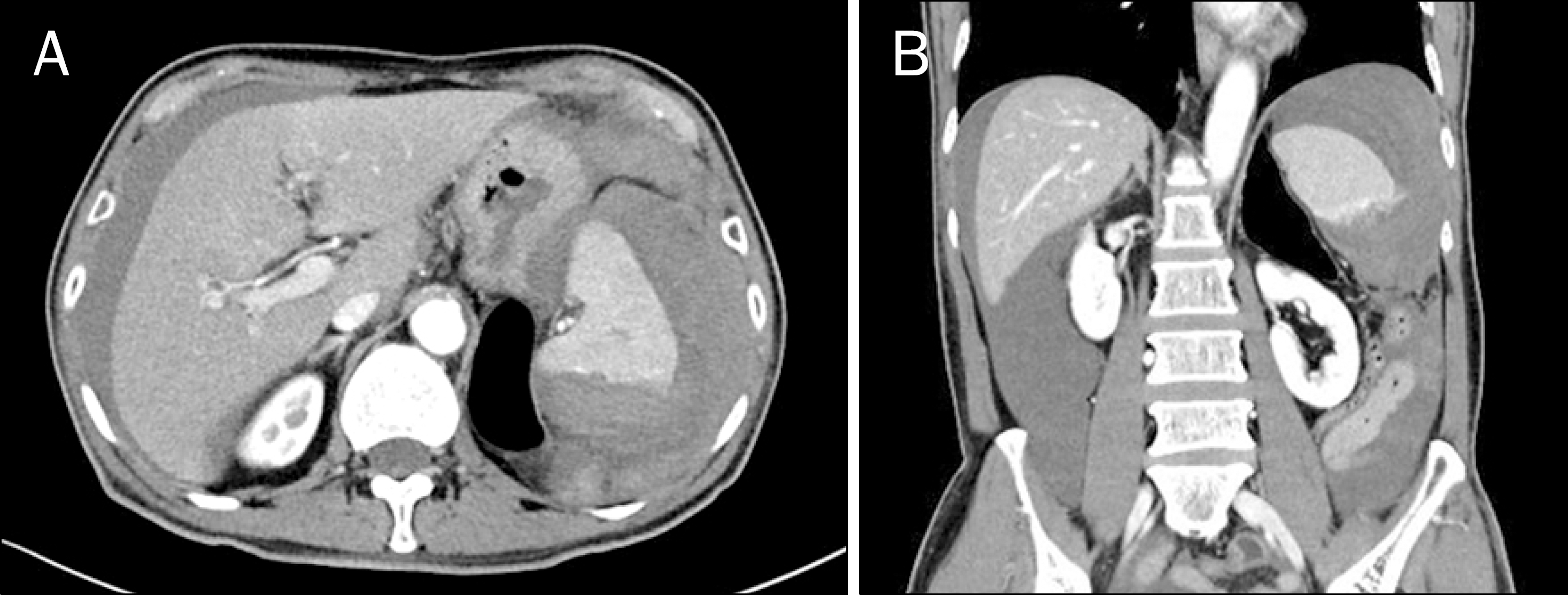
Fig. 2.
Computed tomography scan of the abdomen demonstrated active bleeding from the ruptured spleen (A) with perihepatic-free blood collection (B).

Table 1.
American Association of Surgeons for Trauma Splenic Injury Grading Scale16


 PDF
PDF ePub
ePub Citation
 Print
Print


 XML Download