

 PDF
PDF ePub
ePub Citation
 Print
Print


References
1. Pepys MB. Amyloidosis. Annu Rev Med. 2006; 57:223–241.

2. Okuda Y, Takasugi K, Oyama T, Onuma M, Oyama H. Amyloidosis in rheumatoid arthritis–clinical study of 124 histologically pro-ven cases. Ryumachi. 1994; 34:939–946.
3. Tada S, Iida M, Iwashita A, et al. Endoscopic and biopsy findings of the upper digestive tract in patients with amyloidosis. Gastrointest Endosc. 1990; 36:10–14.
4. Ito T, Sakakibara R, Ito S, et al. Mechanism of constipation in familial amyloid polyneuropathy: a case report. Intern Med. 2006; 45:1173–1175.
5. Mallory A, Struthers JE Jr, Kern F Jr. Persistent hypotension and intestinal infarction in a patient with primary amyloidosis. Gastroenterology. 1975; 68:1587–1592.
6. Jarnum S. Gastrointestinal haemorrhage and protein loss in primary amyloidosis. Gut. 1965; 6:14–18.
7. Chang SS, Lu CL, Tsay SH, Chang FY, Lee SD. Amyloidosis-in-duced gastrointestinal bleeding in a patient with multiple myeloma. J Clin Gastroenterol. 2001; 32:161–163.
8. Ebert EC, Nagar M. Gastrointestinal manifestations of amyloidosis. Am J Gastroenterol. 2008; 103:776–787.
9. Parsons J. Identification of crystalline material from lung biopsy by x-ray diffraction. Henry Ford Hosp Med Bull. 1962; 10:355–357.
10. Maeshima E, Yamada Y, Yukawa S. Massive gastrointestinal hemorrhage in a case of amyloidosis secondary to rheumatoid arthritis. Scand J Rheumatol. 1999; 28:262–264.
11. Mardinger O, Rotenberg L, Chaushu G, Taicher S. Surgical management of macroglossia due to primary amyloidosis. Int J Oral Maxillofac Surg. 1999; 28:129–131.
12. Sattianayagam PT, Hawkins PN, Gillmore JD. Systemic amyloidosis and the gastrointestinal tract. Nat Rev Gastroenterol Hepatol. 2009; 6:608–617.
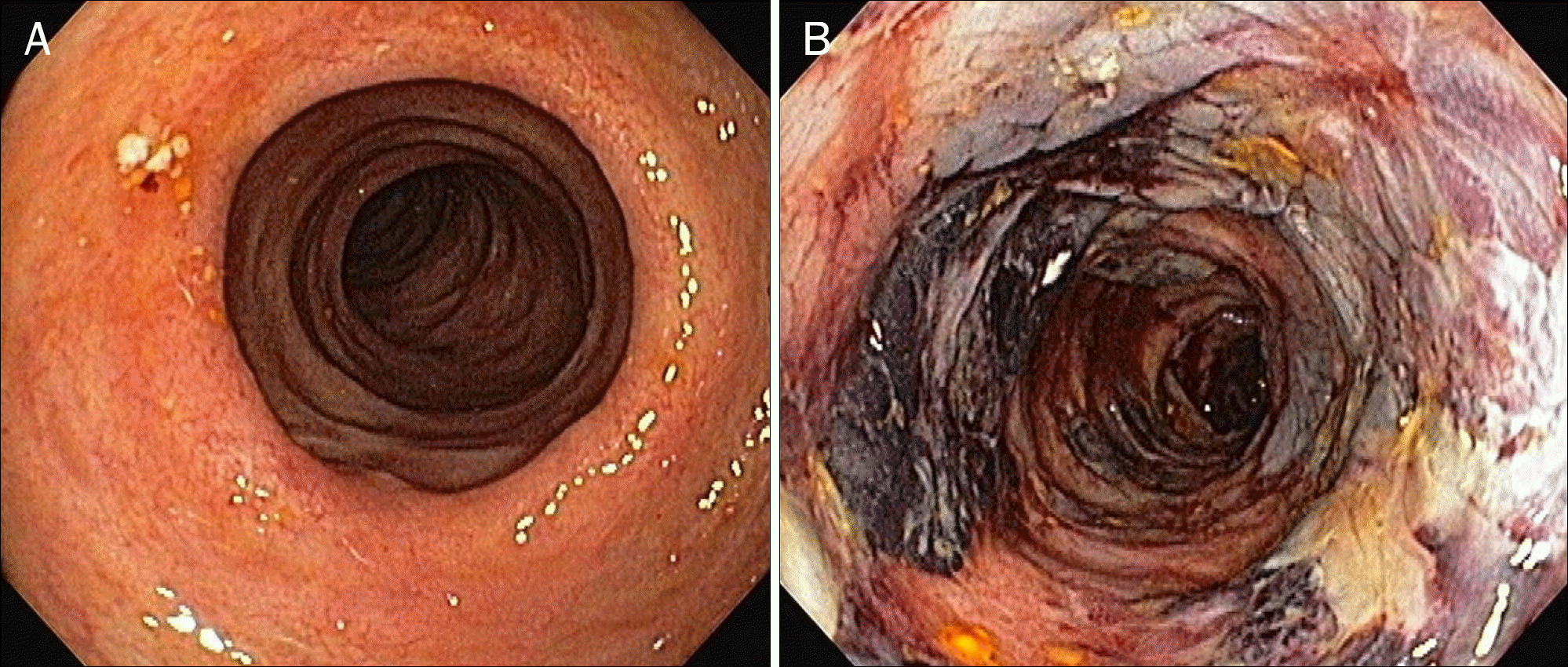
Fig. 1.
Endoscopic findings at initial visit. (A) Coarse mucosa with diminished colonic vasculature is noted throughout the colon. (B) Diffuse mucosal ulceration with necrotic tissue and hemorrhagic bullae are seen on descending and sigmoid colon.
 XML Download