

 PDF
PDF ePub
ePub Citation
 Print
Print


Abstract
Variceal bleeding is common in chronic liver disease and is a frequent cause of acute upper gastrointestinal bleeding. The most common site of varices is the lower oesophagus but they may occur at any location where there are portosystemic anastomoses and collateral vascular formation. Location of ectopic varices at the site of enterocutaneous stomas is rare. We report on three cases of recurrent and severe bleeding from parastomal varices, requiring hospital admission. The patients had chronic liver disease but of different aetiological factors. Variceal formation results from portal hypertension due to chronic liver disease. There are various treatment options for parastomal variceal bleeding, including local, medical, and surgical interventions. Management of parastomal variceal bleeding presents a recurring and difficult problem. Bleeding may be consid-erable and sometimes life threatening. This diagnosis must be considered in patients with chronic liver disease presenting with stomal bleeding, even where the variceal formation may not be readily visible.
References
1. Krige JE, Beckingham IJ. ABC of diseases of liver, pancreas, and biliary system. Portal hypertension-1: varices. BMJ. 2001; 322:348–351.

2. Helmy A, Al Kahtani K, Al Fadda M. Updates in the pathogenesis, diagnosis and management of ectopic varices. Hepatol Int. 2008; 2:322–334.
3. Conte JV, Arcomano TA, Naficy MA, Holt RW. Treatment of bleeding stomal varices. Report of a case and review of the literature. Dis Colon Rectum. 1990; 33:308–314.
4. Norton ID, Andrews JC, Kamath PS. Management of ectopic varices. Hepatology. 1998; 28:1154–1158.
5. Farquharson AL, Bannister JJ, Yates SP. Peristomal varices–life threatening or luminal? Ann R Coll Surg Engl. 2006; 88:W6–W8.
6. Resnick RH, Ishihara A, Chalmers TC, Schimmel EM. A controlled trial of colon bypass in chronic hepatic encephalopathy. Gastroenterology. 1968; 54:1057–1069.
7. Edwards EA. Functional anatomy of the porta-systemic commu-nications. AMA Arch Intern Med. 1951; 88:137–154.
8. Lebrec D, Benhamou JP. Ectopic varices in portal hypertension. Clin Gastroenterol. 1985; 14:105–121.
9. Pennick MO, Artioukh DY. Management of parastomal varices: who re-bleeds and who does not? A systematic review of the literature. Tech Coloproctol. 2013; 17:163–170.
10. Wiesner RH, LaRusso NF, Dozois RR, Beaver SJ. Peristomal varices after proctocolectomy in patients with primary sclerosing cholangitis. Gastroenterology. 1986; 90:316–322.
11. Spier BJ, Fayyad AA, Lucey MR, et al. Bleeding stomal varices: case series and systematic review of the literature. Clin Gastroenterol Hepatol. 2008; 6:346–352.
12. Choi JW, Lee CH, Kim KA, Park CM, Kim JY. Ectopic varices in colonic stoma: MDCT findings. Korean J Radiol. 2006; 7:297–299.
13. Kabeer MA, Jackson L, Widdison AL, Maskell G, Mathew J. Stomal varices: a rare cause of stomal hemorrhage. A report of three cases. Ostomy Wound Manage. 2007; 53:20–22. 24, 26 passim.
14. Wilbur K, Sidhu K. Beta blocker prophylaxis for patients with variceal hemorrhage. J Clin Gastroenterol. 2005; 39:435–440.
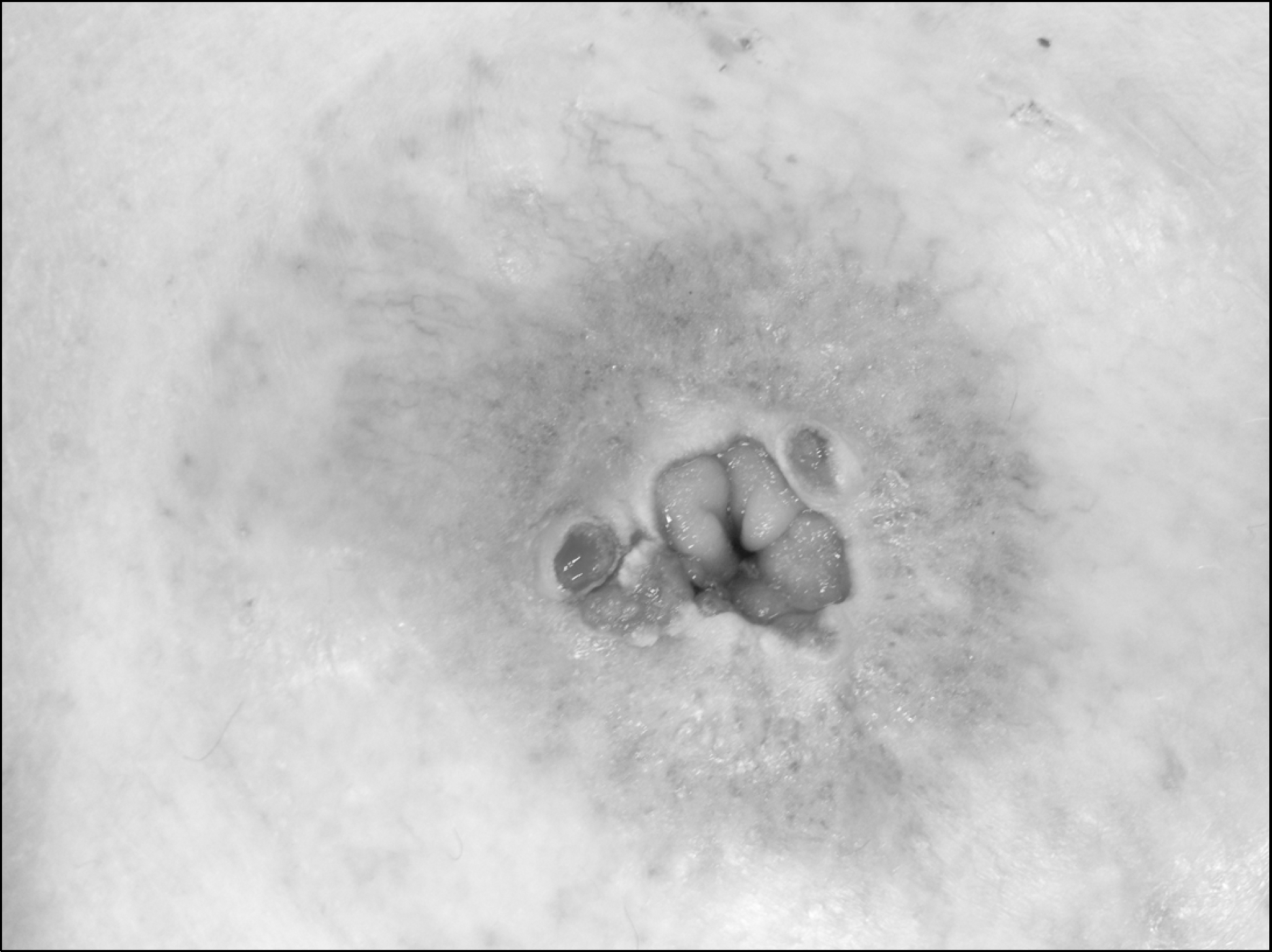
Fig. 1.
Parastomal varices in patient 1. Abnormal radial vascular formation in surrounding skin with extensive circumferential purplish discolouration (raspberry appearance) and ulceration at the mucocutaneous junction of the ileostomy.
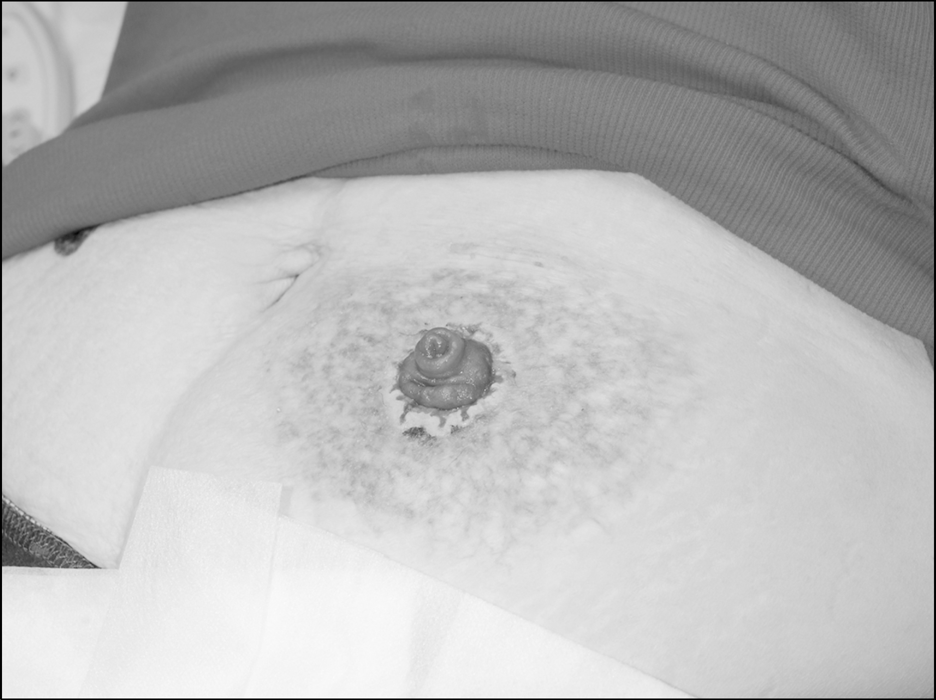
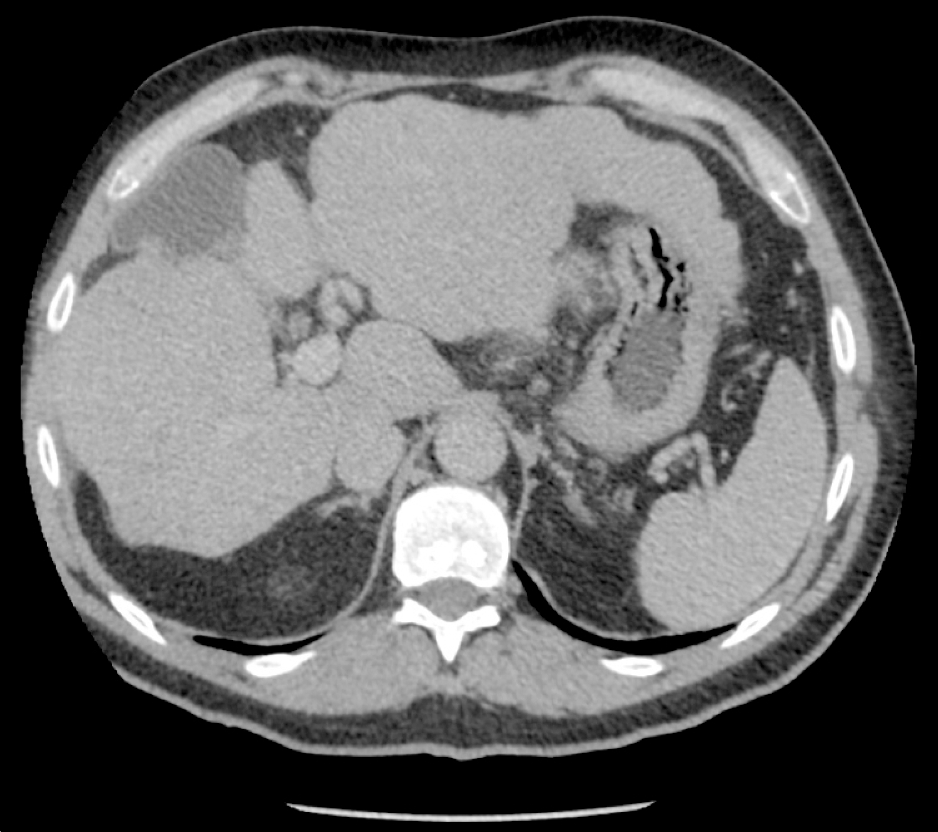
Fig. 2.
CT scan showing a cirrhotic liver due to primary sclerosing cholangitis in patient 2. Irregular and nodular cirrhotic liver with portal hypertension.
 XML Download