

 PDF
PDF ePub
ePub Citation
 Print
Print


References
1. Sashiyama H, Nozawa A, Kimura M, et al. Case report: a case of lymphoepithelioma-like carcinoma of the oesophagus and review of the literature. J Gastroenterol Hepatol. 1999; 14:534–539.

2. MacCarty WC, Mahle AE. Relation of differentiation and lympho-cytic infiltration to postoperative longevity in gastric carcinoma. J Lab Clin Med. 1921; 6:473–480.
3. Watanabe H, Enjoji M, Imai T. Gastric carcinoma with lymphoid stroma. Its morphologic characteristics and prognostic correlations. Cancer. 1976; 38:232–243.
4. Oda K, Tamaru J, Takenouchi T, et al. Association of Epstein-Barr virus with gastric carcinoma with lymphoid stroma. Am J Pathol. 1993; 143:1063–1071.
5. Nakamura S, Ueki T, Yao T, Ueyama T, Tsuneyoshi M. Epstein-Barr virus in gastric carcinoma with lymphoid stroma. Special reference to its detection by the polymerase chain reaction and in situ hybridization in 99 tumors, including a morphologic analysis. Cancer. 1994; 73:2239–2249.
6. Kume T, Oshima K, Yamashita Y, Shirakusa T, Kikuchi M. Relationship between Fas-ligand expression on carcinoma cell and cytotoxic T-lymphocyte response in lymphoepithelioma-like cancer of the stomach. Int J Cancer. 1999; 84:339–343.
7. Hsu DH, de Waal Malefyt R, Fiorentino DF, et al. Expression of in-terleukin-10 activity by Epstein-Barr virus protein BCRF1. Science. 1990; 250:830–832.
8. Endo T, Okuda H, Arimura Y, et al. A case of early gastric carcinoma with lymphoid stroma: diagnostic usefulness of endosono-graphy. Dig Endosc. 1998; 10:240–243.
9. Lee JY, Kim KM, Min BH, Lee JH, Rhee PL, Kim JJ. Epstein-Barr virus-associated lymphoepithelioma-like early gastric carcinomas and endoscopic submucosal dissection: case series. World J Gastroenterol. 2014; 20:1365–1370.
10. Tak DH, Jeong HY, Seong JK, Moon HS, Kang SH. Comparison of clinical characteristics and prognostic factors between gastric lymphoepithelioma-like carcinoma and gastric adenocarcinoma. Korean J Gastroenterol. 2013; 62:272–277.
Fig. 1.
Endoscopic finding. About 2.0 cm sized protruding lesion with surface erosion and bridging folds is seen on the less curvature of low body.
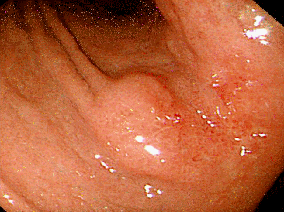
Fig. 2.
EUS finding. EUS shows 2.0 cm sized hypoecohoic mass involving mainly the third layer and focal fourth layer of gastric wall.
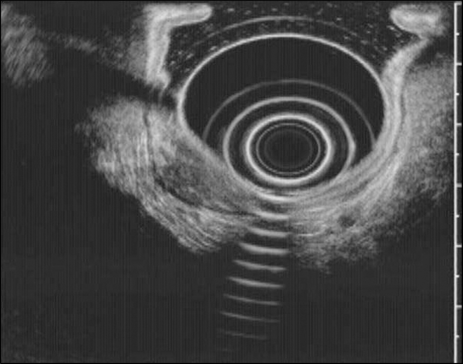
Fig. 3.
Endoscopic findings during endoscopic resection. (A) Whitish mass lesion is revealed after mucosa denudation. (B) The mass lesion is shown to have invaded the proper muscle layer.
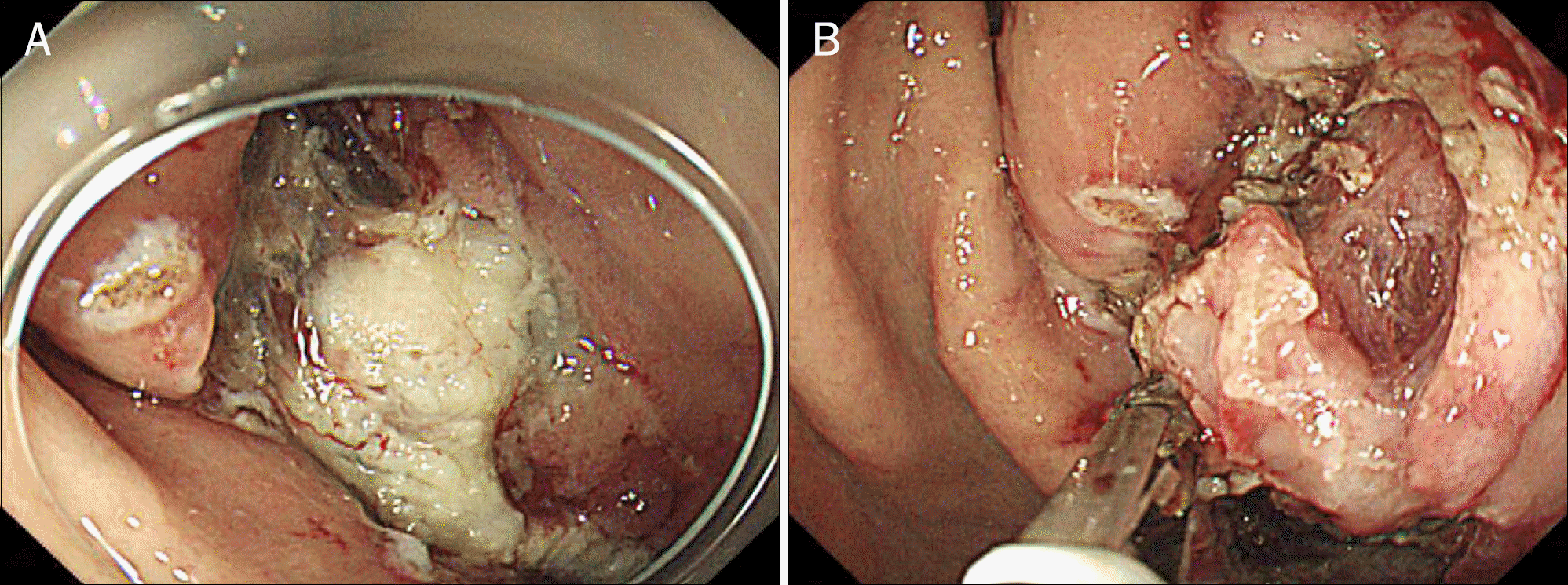
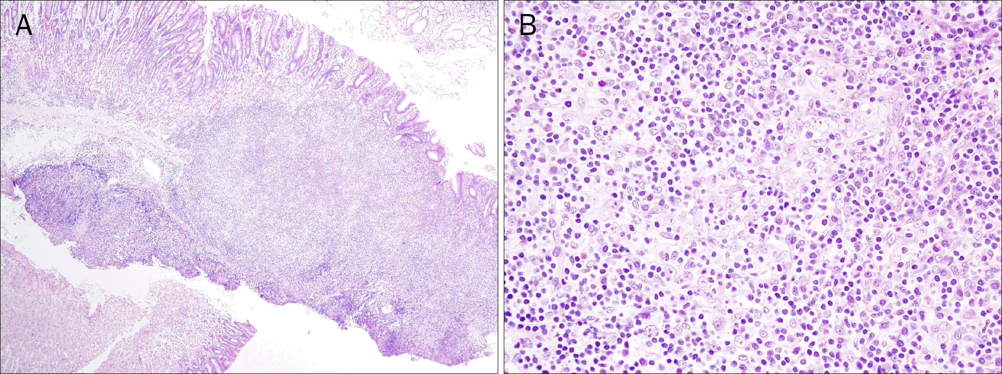
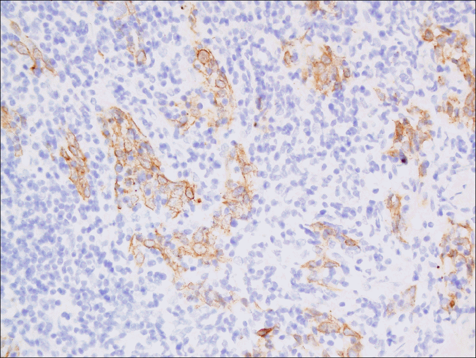
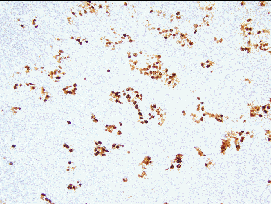
Fig. 4.
Histologic findings (H&E). (A) The submucosal tumor is well demarcated (×40). (B) Large sized atypical cells are seen with many lymphocytes on the background (×400).
 XML Download