Abstract
Hepatic sinusoidal dilatation is a rare benign vascular disorder characterized by focal dilatation of the sinusoidal spaces. In most cases, the underlying etiology is unclear but it may be related to the impairment of venous outflow or sinusoidal infiltration by diverse causes. Diagnosing hepatic sinusoidal dilatation based soley on imaging study is not easy since there are no pathognomonic radiologic findings indicative of this condition. Recently, the authors experience two cases of hepatic sinusoidal infiltration. The first patient was a 53-year-old man detected to have multiple hepatic nodules on ultrasonography (US) during a routine medical checkup. The second patient was an 82-year-old woman with abdominal discomfort who was referred from local clinic with high suspicion of hepatic metastases on US. In both cases, CT scan demonstrated multiple nodules with rim enhancement on arterial phase that became isodense to adjacent liver parenchyma on delayed phase. On MRI, these nodules showed rim enhancement on arterial phase, had high signal intensity on T2 weighted images, and became iso-intense with partial defect on hepatobiliary phase. Because imaging studies could not exclude the presence of hepatic metastases, liver biopsy was performed and it demonstrated hepatic sinusoidal dilatation with well preserved reticulin fiber without any evidence of malignancy. Herein, we report two cases of idiopathic hepatic sinusoidal dilatation mimicking hepatic metastases.
Go to : 
References
1. Degott C, Potet F. Peliosis hepatis and sinusoidal dilatatio. Arch Anat Cytol Pathol. 1984; 32:296–300.
2. Winkler K, Poulsen H. Liver disease with periportal sinusoidal dilatation. A possible complication to contraceptive steroids. Scand J Gastroenterol. 1975; 10:699–704.
3. Kakar S, Kamath PS, Burgart LJ. Sinusoidal dilatation and con-gestion in liver biopsy: is it always due to venous outflow impair-ment? Arch Pathol Lab Med. 2004; 128:901–904.

4. Zhou H, Wang YX, Lou HY, Xu XJ, Zhang MM. Hepatic sinusoidal obstruction syndrome caused by herbal medicine: CT and MRI features. Korean J Radiol. 2014; 15:218–225.

5. Helmy A. Review article: updates in the pathogenesis and therapy of hepatic sinusoidal obstruction syndrome. Aliment Pharmacol Ther. 2006; 23:11–25.

6. DeLeve LD, Valla DC, Garcia-Tsao G. American Association for the Study Liver Diseases. Vascular disorders of the liver. Hepatology. 2009; 49:1729–1764.

7. Laffón A, Moreno A, Gutierrez-Bucero A, Ossorio C, Sabando P, Moreno-Otero R. Hepatic sinusoidal dilatation in rheumatoid arthritis. J Clin Gastroenterol. 1989; 11:653–657.

8. Bruguera M, Caballero T, Carreras E, Aymerich M, Rodés J, Rozman C. Hepatic sinusoidal dilatation in Hodgkin's disease. Liver. 1987; 7:76–80.

9. Capron JP, Lemay JL, Gontier MF, Dupas JL, Capron-Chivrac D, Lorriaux A. Hepatic sinusoidal dilatation in Crohn's disease. Scand J Gastroenterol. 1979; 14:987–992.
10. Curciarello J, Castelletto R, Barbero R, et al. Hepatic sinusoidal dilatation associated to giant lymph node hyperplasia (Castle-man's): a new case in a patient with periorbital xanthelasmas and history of celiac disease. J Clin Gastroenterol. 1998; 27:76–78.
11. Fisher MR, Neiman HL. Periportal sinusoidal dilatation associated with pregnancy. Cardiovasc Intervent Radiol. 1984; 7:299–302.

12. Balázs M. Sinusoidal dilatation of the liver in patients on oral contraceptives. Electron microscopical study of 14 cases. Exp Pathol. 1988; 35:231–237.

13. Weinberger M, Garty M, Cohen M, Russo Y, Rosenfeld JB. Ultrasonography in the diagnosis and follow-up of hepatic sinusoidal dilatation. Arch Intern Med. 1985; 145:927–929.

14. Uchino K, Fujisawa M, Watanabe T, et al. Oxaliplatin-induced liver injury mimicking metastatic tumor on images: a case report. Jpn J Clin Oncol. 2013; 43:1034–1038.

15. Agostini J, Benoist S, Seman M, et al. Identification of molecular pathways involved in oxaliplatin-associated sinusoidal dilatation. J Hepatol. 2012; 56:869–876.

16. Gerlag PG, van Hooff JP. Hepatic sinusoidal dilatation with portal hypertension during azathioprine treatment: a cause of chronic liver disease after kidney transplantation. Transplant Proc. 1987; 19:3699–3703.
17. Bruguera M, Aranguibel F, Ros E, Rodés J. Incidence and clinical significance of sinusoidal dilatation in liver biopsies. Gastroenterology. 1978; 75:474–478.

Go to : 
 | Fig. 1.Liver dynamic CT findings of two cases; a 53-year-old man (A-C) and an 82-year-old woman (D-F). In the first case, CT scan shows multiple variable sized nodules (arrows) with rim enhancement on arterial phase (A), central enhancement (arrowheads) on portal phase (B) and isodense on delayed phase (C). In the second case, CT scan shows multiple variable sized nodules (arrows) with rim enhancement on arterial phase (D) which become isodense on portal (E) and delayed phase (F). |
 | Fig. 2.MRI findings of two cases; a 53-year-old man (A-D) and an 82-year-old woman (E-G). Multiple variable sized nodules (black arrows) show rim enhancement on arterial phase (A, E), have iso-signal intensity on venous phase (B, F), demonstrate high signal intensity (white arrows) on T2 weighted image (C, G), and become iso-signal intense with partial defect (white arrowheads) on hepatobiliary phase (D, H). |
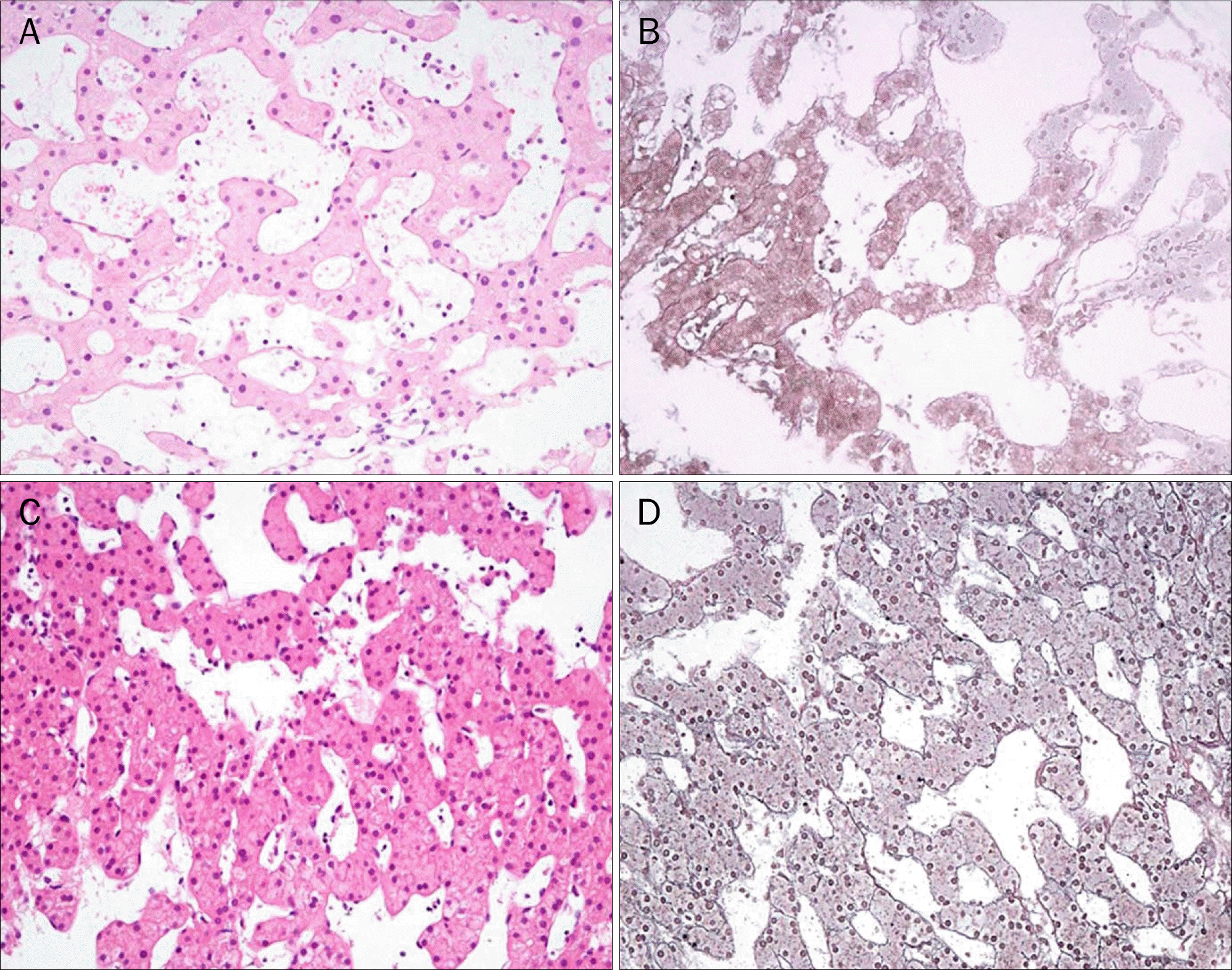
 | Fig. 3.Pathologic findings of two cases. In the first case (a 53-year-old man), (A) sinusoids are dilated, which indicates hepatic sinusoidal dilatation, but malignant cells are not present (H&E, ×200). (B) Reticulin networks are well preserved without disruption, which rules out peliosis hepatis (Reticulin stain, ×200). In the second case (an 82-year-old woman), normal hepatocyte and dilated sinusoidal space are noted similar to the previous case (C: H&E, ×200; D: Reticulin stain, ×200). |


 PDF
PDF ePub
ePub Citation
 Print
Print


 XML Download