Abstract
Overtube provides a conduit for the passage of endoscope into the digestive tract. Esophageal perforation with mediastinitis is a rare overtube-related complication. Until now, no reports have been published regarding the esophageal perforation which developed many months after the original procedure using the overtube. A 56-year-old female visited our hospital complaining of chest pain and back pain that began 14 days ago. The patient underwent esophageal variceal ligation using the overtube 12 months earlier. She was diagnosed with esophageal perforation with mediastinitis which extended to intervertebral and epidural space. The cause of this condition was considered to have been related to the use of overtube. Management of delayed perforation remains controversial. Although surgical management might be the preferred mode of treatment, she underwent local N-butyl 2-cyanoacrylate injection therapy and temporary stent therapy with antibiotics due to high operative risk. Herein, we report a case of overtube-related delayed esophageal perforation with mediastinitis that was successfully treated by nonoperative management.
Go to : 
References
1. Schmitz RJ, Sharma P, Badr AS, Qamar MT, Weston AP. Incidence and management of esophageal stricture formation, ulcer bleeding, perforation, and massive hematoma formation from sclerotherapy versus band ligation. Am J Gastroenterol. 2001; 96:437–441.

2. Goldschmiedt M, Haber G, Kandel G, Kortan P, Marcon N. A safe-ty maneuver for placing overtubes during endoscopic variceal ligation. Gastrointest Endosc. 1992; 38:399–400.

3. Lindenmann J, Matzi V, Neuboeck N, et al. Management of esophageal perforation in 120 consecutive patients: clinical impact of a structured treatment algorithm. J Gastrointest Surg. 2013; 17:1036–1043.

4. Carrott PW Jr, Low DE. Advances in the management of esophageal perforation. Thorac Surg Clin. 2011; 21:541–555.

5. Wells CD, Fleischer DE. Overtubes in gastrointestinal endoscopy. Am J Gastroenterol. 2008; 103:745–752.

6. Dinning JP, Jaffe PE. Delayed presentation of esophageal perforation as a result of overtube placement. J Clin Gastroenterol. 1997; 24:250–252.

7. Michel L, Grillo HC, Malt RA. Operative and nonoperative management of esophageal perforations. Ann Surg. 1981; 194:57–63.

8. Hasan S, Jilaihawi AN, Prakash D. Conservative management of iatrogenic oesophageal perforations–a viable option. Eur J Car-diothorac Surg. 2005; 28:7–10.
9. Brinster CJ, Singhal S, Lee L, Marshall MB, Kaiser LR, Kucharczuk JC. Evolving options in the management of esophageal perforation. Ann Thorac Surg. 2004; 77:1475–1483.

10. Kim HG, Cho JW, Park SJ, et al. Two cases of alimentary tract fistula treated by endoscopic local injection therapy. Korean J Gastrointest Endosc. 2003; 26:426–430.
11. van Heel NC, Haringsma J, Spaander MC, Bruno MJ, Kuipers EJ. Short-term esophageal stenting in the management of benign perforations. Am J Gastroenterol. 2010; 105:1515–1520.

12. Radecke K, Gerken G, Treichel U. Impact of a self-expanding, plastic esophageal stent on various esophageal stenoses, fistu- las, and leakages: a single-center experience in 39 patients. Gastrointest Endosc. 2005; 61:812–818.
13. Langer FB, Wenzl E, Prager G, et al. Management of post-operative esophageal leaks with the Polyflex self-expanding cov-ered plastic stent. Ann Thorac Surg. 2005; 79:398–403.

Go to : 
 | Fig. 1.Endoscopic findings. (A) Endoscopic elastic band ligation for active variceal hemorrhage was performed 12 months ago. (B) Follow-up endoscopy at 3 month after the procedure revealed small esophageal diverticulum at the ligation site. (C) Follow-up endoscopic finding at admission showed esophageal fistula (arrowhead) on mid-esophagus. |
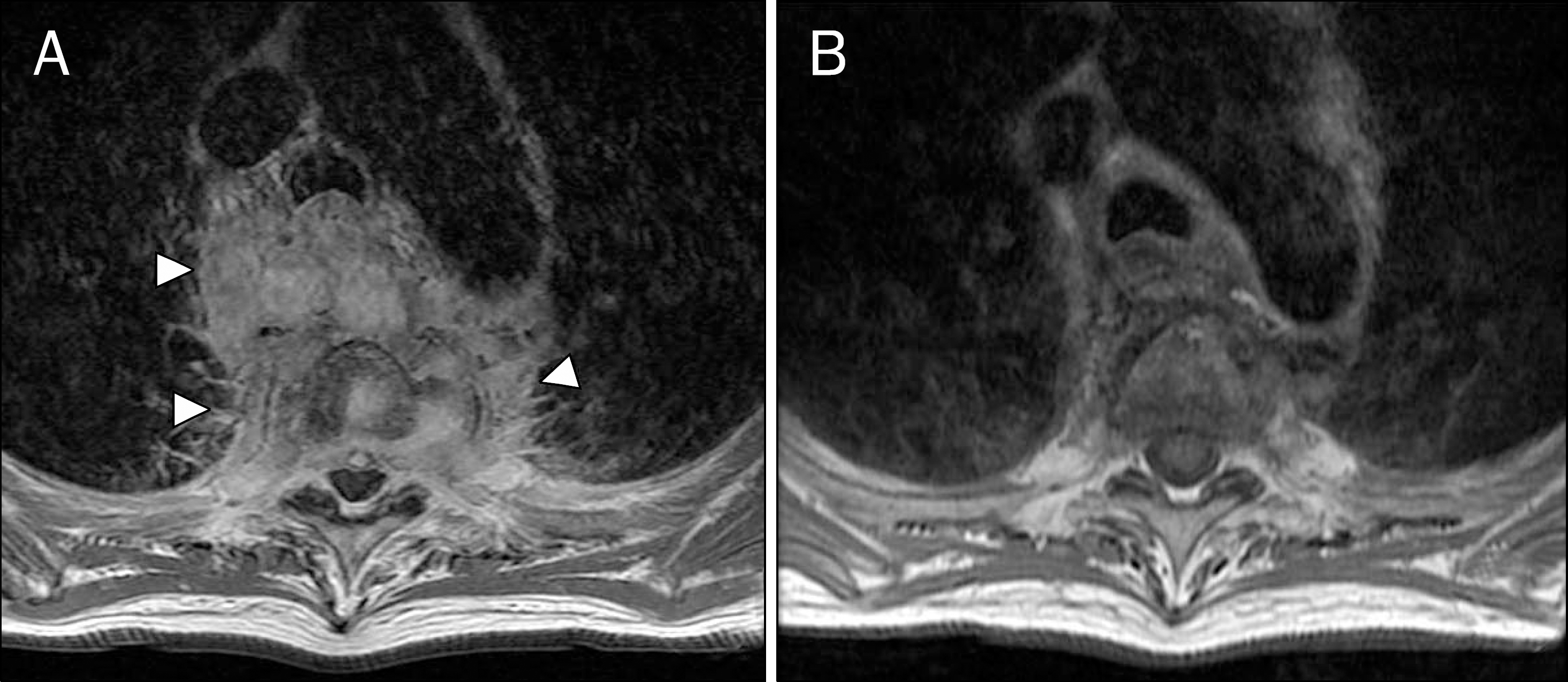
 | Fig. 2.MRI findings. (A) Posterior mediastinal inflammatory mass that extends to intervertebral and epidural space can be seen (arrowheads). (B) Follow-up MRI taken after 4 months of treatment shows markedly decreased extent of posterior mediastinal inflammatory lesion. |


 PDF
PDF ePub
ePub Citation
 Print
Print


 XML Download