Abstract
Gallbladder tuberculosis is an extremely rare disease that is rarely reported in the literature. Arriving at the correct diagnosis of gallbladder tuberculosis is difficult, and it is usually made by histopathologic examination after cholecystectomy. However, due to the low sensitivity of acid-fast stain and culture result, diagnosing gallbladder tuberculosis is still demanding even after tissue acquisition. To overcome this problem, tuberculosis-polymerase chain reaction (TB-PCR) is performed on the resected specimen, which has high sensitivity and specificity. A 70-year-old female who had previously undergone total gastrectomy for advanced gastric cancer was admitted with right upper quadrant pain. Abdominal ultrasonography and computed tomography revealed acute cholecystitis without gallstones or sludge. She underwent cholecystectomy and the histopathologic finding of the specimen showed chronic active cholecystitis without gallstones or sludge. Because she was suspected to have pulmonary tuberculosis, TB-PCR was also performed on the resected gallbladder. TB-PCR showed positive reaction for Mycobacterium tuberculosis and we could diagnose it as gallbladder tuberculosis. Herein, we present a case of gallbladder tuberculosis diagnosed by TB-PCR from resected gallbladder.
References
1. Yu R, Liu Y. Gallbladder tuberculosis: case report. Chin Med J (Engl). 2002; 115:1259–1261.
2. Hahn ST, Park SH, Shin WS, Kim CY, Shinn KS. Gallbladder tuberculosis with perforation and intrahepatic biloma. J Clin Gastroenterol. 1995; 20:84–86.
3. Hyun JH, Song CW, Ryu HS, et al. A case of gallbladder tuberculosis. Korean J Gastroenterol. 1998; 31:383–386.
4. Hulnick DH, Megibow AJ, Naidich DP, Hilton S, Cho KC, Balthazar EJ. Abdominal tuberculosis: CT evaluation. Radiology. 1985; 157:199–204.

5. Saluja SS, Ray S, Pal S, et al. Hepatobiliary and pancreatic tuberculosis: a two decade experience. BMC Surg. 2007; 7:10.

6. Abu-Zidan FM, Zayat I. Gallbladder tuberculosis (case report and review of the literature). Hepatogastroenterology. 1999; 46:2804–2806.
8. Jain R, Sawhney S, Bhargava D, Berry M. Gallbladder tuberculosis: sonographic appearance. J Clin Ultrasound. 1995; 23:327–329.

9. Xu XF, Yu RS, Qiu LL, Shen J, Dong F, Chen Y. Gallbladder tuberculosis: CT findings with histopathologic correlation. Korean J Radiol. 2011; 12:196–202.

10. Rouas L, Mansouri F, Jahid A, et al. Gallbladder tuberculosis associated with cholelithiasis. Rev Med Liege. 2003; 58:757–760.
11. Ramia JM, Muffak K, Fernández A, Villar J, Garrote D, Ferron JA. Gallbladder tuberculosis: false-positive PET diagnosis of gallbladder cancer. World J Gastroenterol. 2006; 12:6559–6560.

12. Chawla K, Gupta S, Mukhopadhyay C, Rao PS, Bhat SS. PCR for M. tuberculosis in tissue samples. J Infect Dev Ctries. 2009; 3:83–87.

13. Salian NV, Rish JA, Eisenach KD, Cave MD, Bates JH. Polymerase chain reaction to detect Mycobacterium tuberculosis in histologic specimens. Am J Respir Crit Care Med. 1998; 158:1150–1155.
14. Yun EY, Cho SH, Go SI, et al. Usefulness of real-time PCR to detect Mycobacterium tuberculosis and nontuberculous mycobacteria. Tuberc Respir Dis. 2010; 69:250–255.
15. García-Elorriaga G, Gracida-Osorno C, Carrillo-Montes G, González- Bonilla C. Clinical usefulness of the nested polymerase chain reaction in the diagnosis of extrapulmonary tuberculosis. Salud Publica Mex. 2009; 51:240–245.

16. Joint Committee for the Development of Korean Guidelines for Tuberculosis. Korean guidelines for tuberculosis. 1st ed.Chungbuk: Korea Centers for Disease Control and Prevention;2011.
17. Blumberg HM, Burman WJ, Chaisson RE, et al. American Thoracic Society, Centers for Disease Control and Prevention and the Infectious Diseases Society. American Thoracic Society/Centers for Disease Control and Prevention/Infectious Diseases Society of America: treatment of tuberculosis. Am J Respir Crit Care Med. 2003; 167:603–662.
18. World Health Organization. Treatment of tuberculosis: guidelines for national programmes. 4th ed.Geneva: World Health Organization;2010.
Fig. 1.
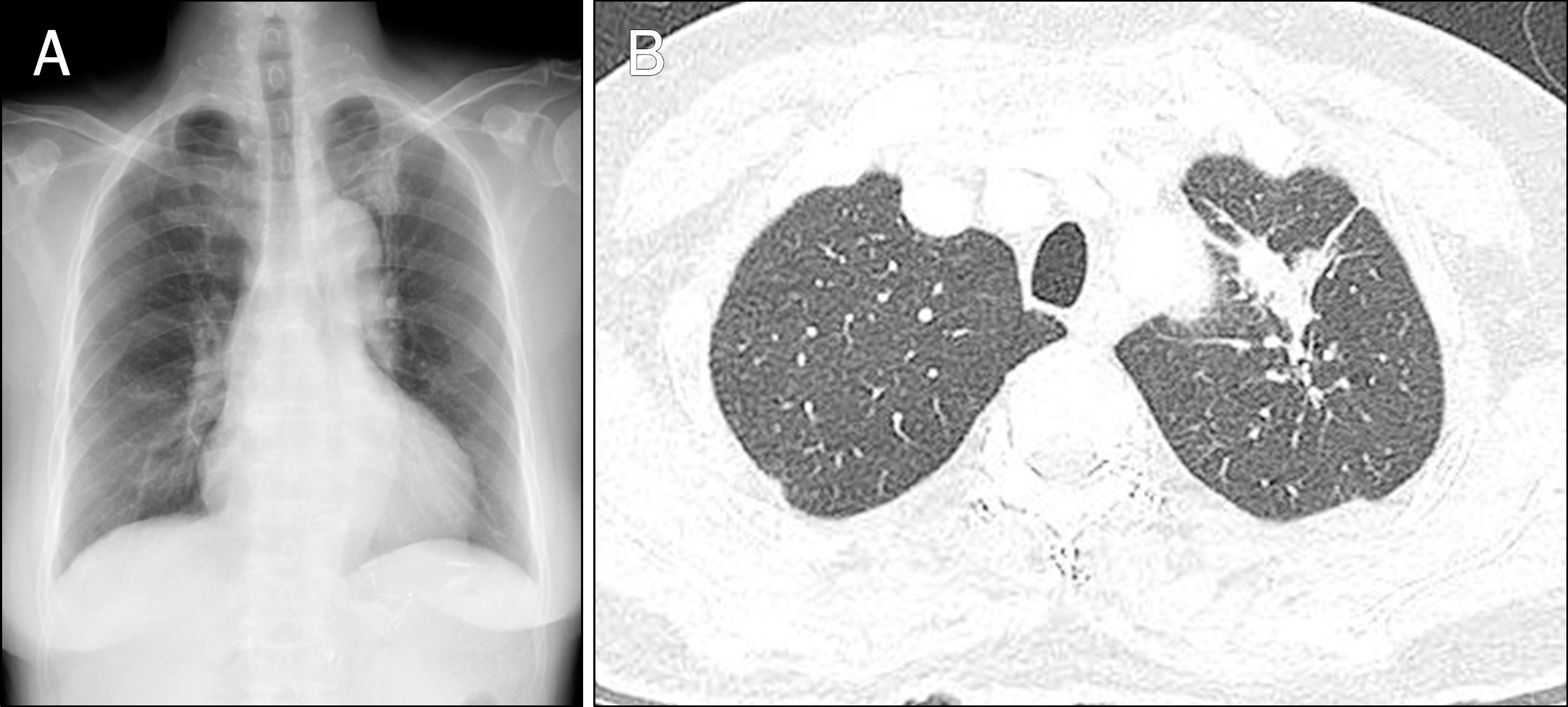
(A) Chest X-ray shows increased nodular opacity in left upper lobe. (B) Chest CT reveals a 4-cm-sized irregular low attenuated consolidative lesion in left upper lobe.

Fig. 2.
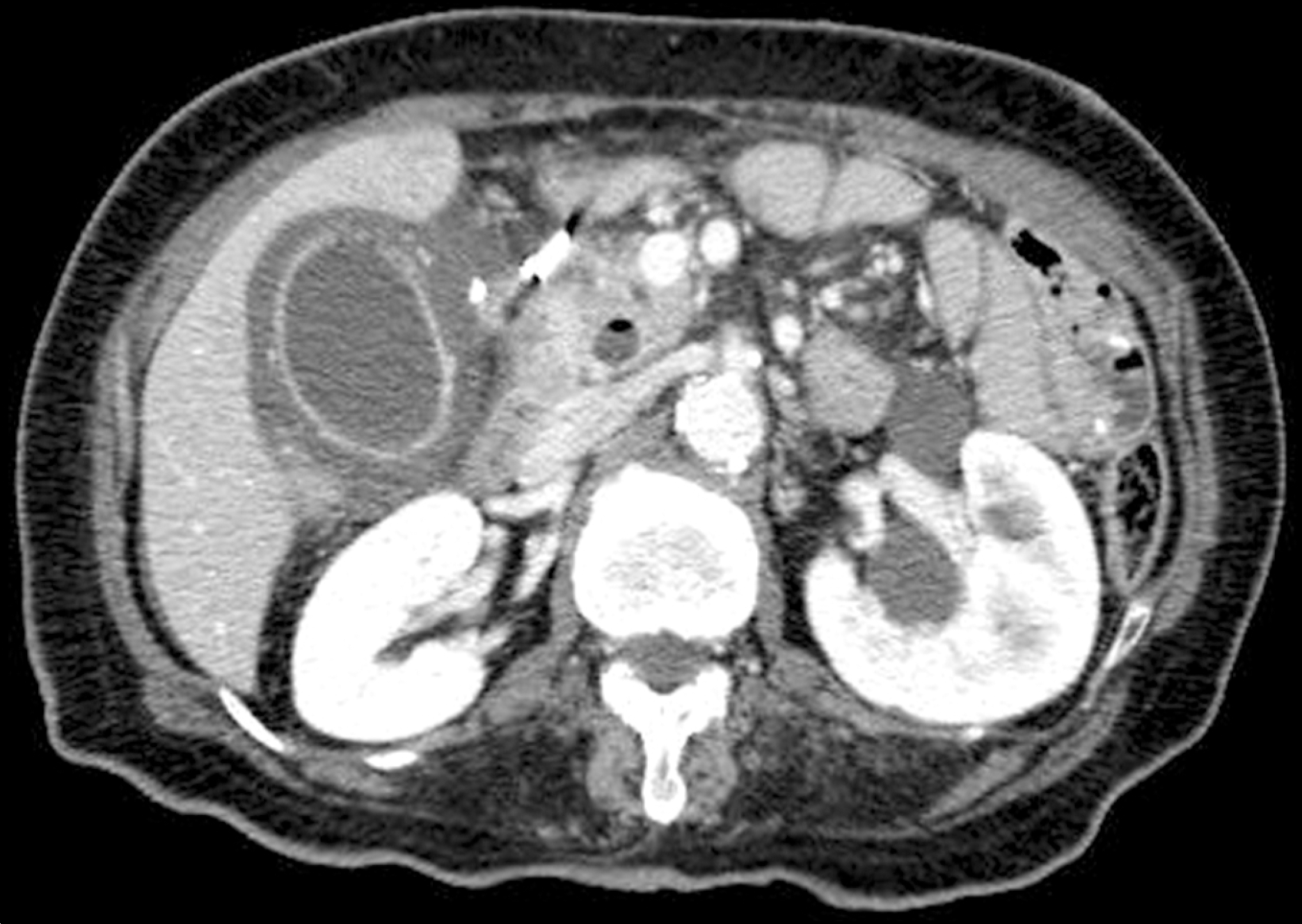
Abdominal CT shows distended gallbladder with diffuse gallbladder wall thickening, hyperemic change of adjacent liver parenchyma, and extrahepatic duct dilatation. The CT scan also shows mild swelling of pancreas head and peripancreatic fat infiltration.

Fig. 4.
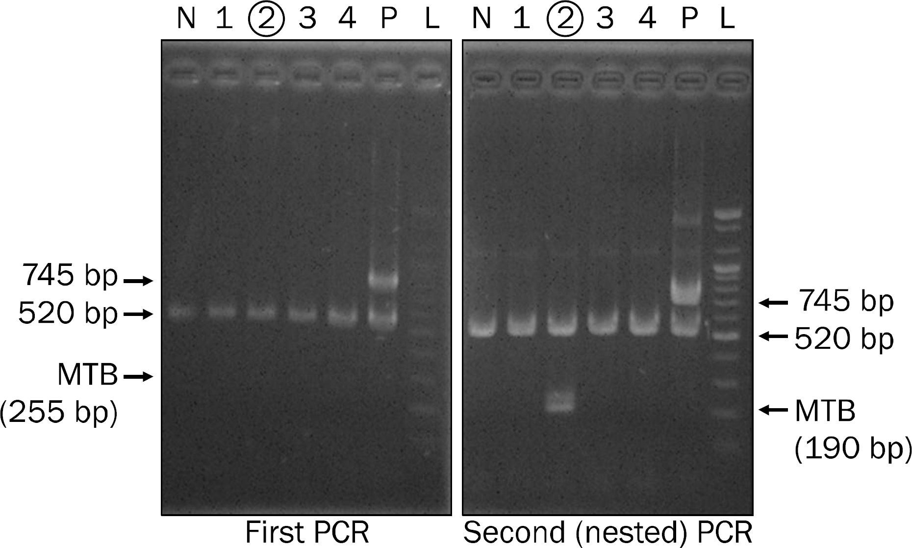
Nested-PCR for Mycobacterium tuberculosis. To eliminate any possibility of cross contamination from M. tuberculosis (MTB) positive control PCR, amplicon size 745 bp of the positive control PCR was desinged. The internal control is 520 bp in the first and second (nested) PCR. Normally, the first PCR product (255 bp) does not show up but could very rarely be generated in the presence of high titer M. tuberculosis. In our patient (lane 2), 255 bp band is also not detected on first PCR. However, the second (nested) PCR assay shows positive reaction for M. tuberculosis (190 bp). Lanes 1, 3, 4 are samples from other patients. N, negative control lane; P, positive control lane; L, ladder (molecular weight marker).


 PDF
PDF ePub
ePub Citation
 Print
Print


 XML Download