

 PDF
PDF ePub
ePub Citation
 Print
Print


Abstract
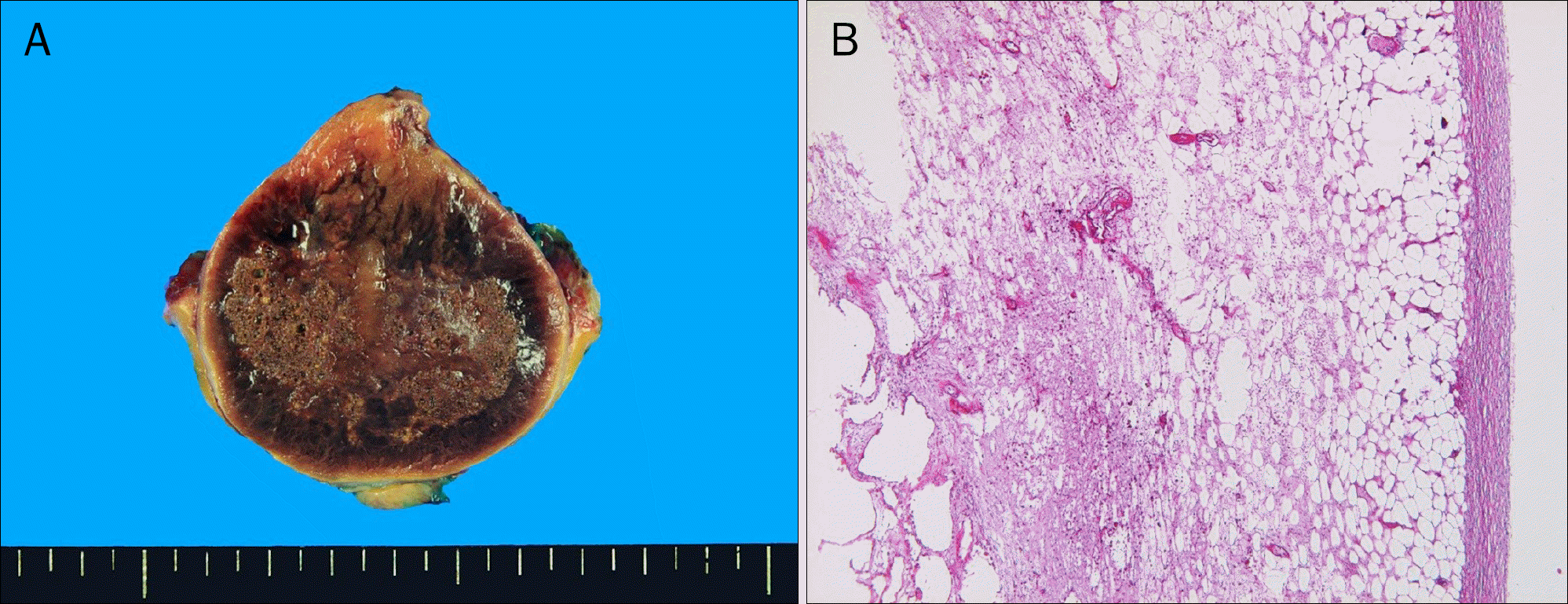
Lipomas are common benign tumors of mature adipose tissue, enclosed by thin fibrous capsules. They can occur on any part of the body; however, peritoneal lipoma is extremely rare. We encountered a case of a 75-year-old man presenting with intermittent abdominal pain, who had undergone right hemicolectomy due to colon cancer. Abdominal computerized tomography showed a well-defined heterogenous fatty mass measuring 4.5×3.5 cm in size, suggesting fat necrosis located in the abdominal wall. Laparotomy showed a very large soft mass of peritoneum. Pathologically, the tumor was diagnosed as lipoma containing fat necrosis located in parietal peritoneum not fixed to any organs, but with small bowel adhesion. Due to its rare etiologic origin and obscure cause of development, we report on a case of lipoma of parietal peritoneum causing abdominal pain.
Go to : 
References
1. Piattelli A, Fioroni M, Iezzi G, Rubini C. Osteolipoma of the tongue. Oral Oncol. 2001; 37:468–470.

2. Thompson WM, Kende AI, Levy AD. Imaging characteristics of gastric lipomas in 16 adult and pediatric patients. AJR Am J Roentgenol. 2003; 181:981–985.
3. Rapidis AD. Lipoma of the oral cavity. Int J Oral Surg. 1982; 11:30–35.
4. Pfeil SA, Weaver MG, Abdul-Karim FW, Yang P. Colonic lipomas: outcome of endoscopic removal. Gastrointest Endosc. 1990; 36:435–438.
5. Tascilar O, Cakmak GK, Gün BD, et al. Clinical evaluation of submucosal colonic lipomas: decision making. World J Gastroenterol. 2006; 12:5075–5077.
6. Tamura S, Yokoyama Y, Morita T, Tadokoro T, Higashidani Y, Onishi S. "Giant" colon lipoma: what kind of findings are necessary for the indication of endoscopic resection? Am J Gastroenterol. 2001; 96:1944–1946.
7. Singaporewalla RM, Thamboo TP, Rauff A, Cheah WK, Mukherjee JJ. Acute abdominal pain secondary to retroperitoneal bleeding from a giant adrenal lipoma with review of literature. Asian J Surg. 2009; 32:172–176.
8. Wolko JD, Rosenfeld DL, Lazar MJ, Underberg-Davis SJ. Torsion of a giant mesenteric lipoma. Pediatr Radiol. 2003; 33:34–36.
9. Ilhan H, Tokar B, Iş iksoy S, Koku N, Pasaoglu O. Giant mesenteric lipoma. J Pediatr Surg. 1999; 34:639–640.
10. Barut I, Tarhan OR, Cerci C, Ciris M, Tasliyar E. Lipoma of the pari-etal peritoneum: an unusual cause of abdominal pain. Ann Saudi Med. 2006; 26:388–390.
11. de Visscher JG. Lipomas and fibrolipomas of the oral cavity. J Maxillofac Surg. 1982; 10:177–181.
12. Levy AD, Arnáiz J, Shaw JC, Sobin LH. From the archives of the AFIP: primary peritoneal tumors: imaging features with pathologic correlation. Radiographics. 2008; 28:583–607.
13. Pickhardt PJ, Bhalla S. Primary neoplasms of peritoneal and sub-peritoneal origin: CT findings. Radiographics. 2005; 25:983–995.
14. Weinberg T, Feldman M Sr. Lipomas of the gastrointestinal tract. Am J Clin Pathol. 1955; 25:272–281.
15. Haller JD, Roberts TW. Lipomas of the colon: a clinicopathologic study of 20 cases. Surgery. 1964; 55:773–781.
16. Signorini M, Campiglio GL. Posttraumatic lipomas: where do they really come from? Plast Reconstr Surg. 1998; 101:699–705.
17. Moretti-Ferreira D, Koiffmann CP, Souza DH, Diament AJ, Wajntal A. Macrocephaly, multiple lipomas, and hemangiomata (Bannayan-Zonana syndrome): genetic heterogeneity or autosomal dominant locus with at least two different allelic forms? Am J Med Genet. 1989; 34:548–551.
18. Zhang H, Erickson-Johnson M, Wang X, et al. Molecular testing for lipomatous tumors: critical analysis and test recommendations based on the analysis of 405 extremity-based tumors. Am J Surg Pathol. 2010; 34:1304–1311.
Go to :

 XML Download