

 PDF
PDF ePub
ePub Citation
 Print
Print


Abstract
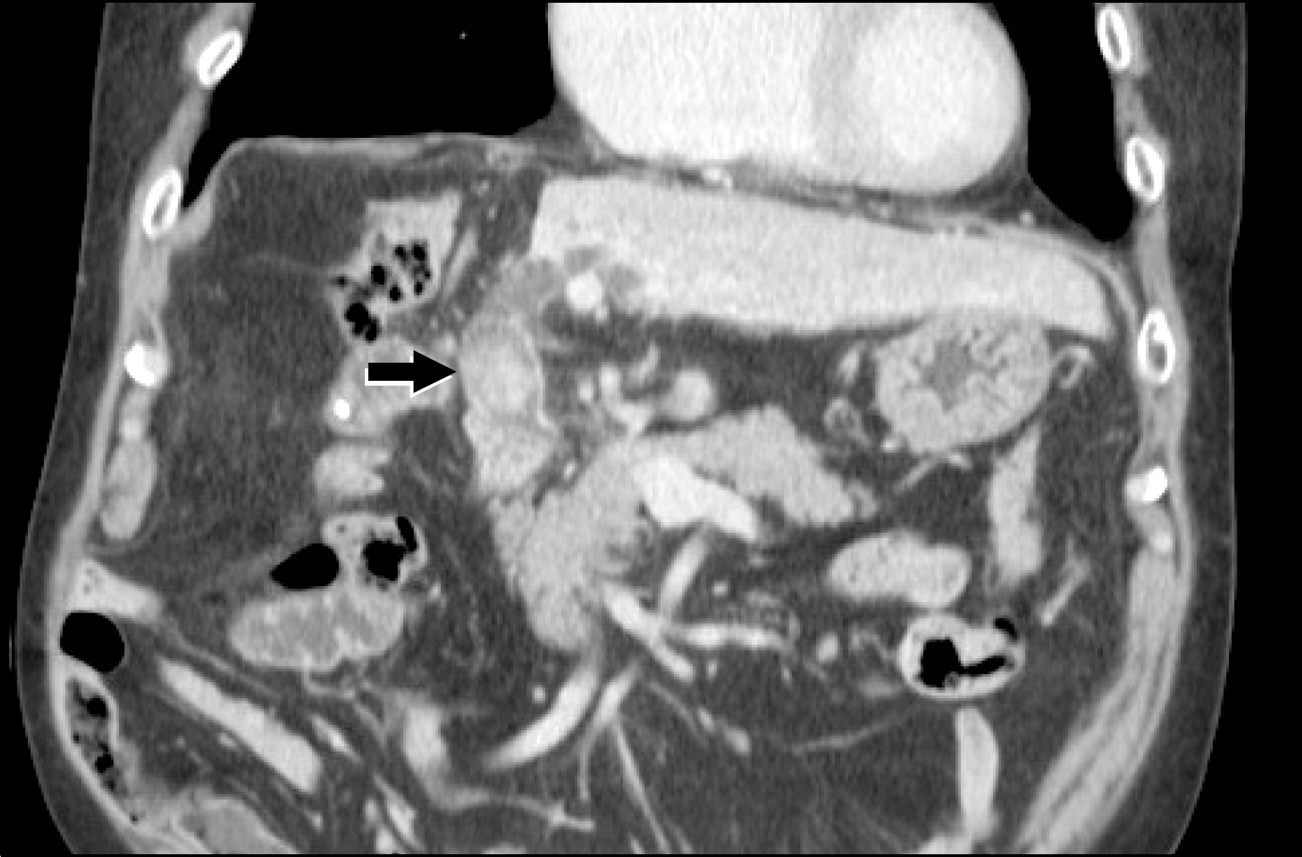
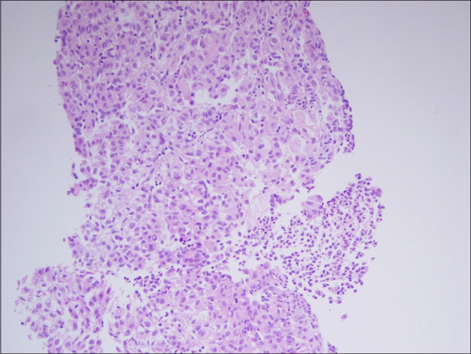
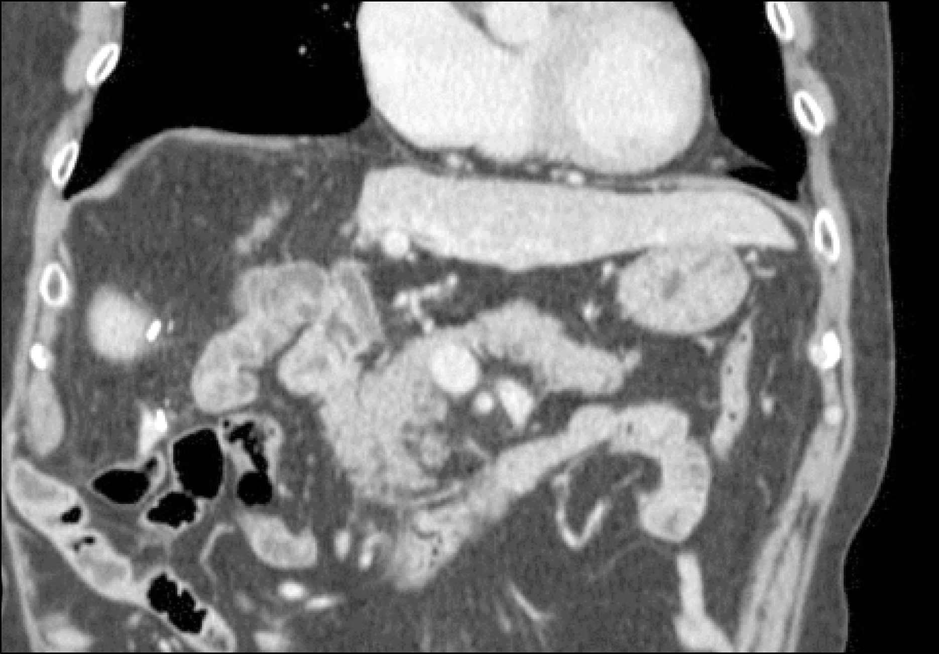
Invasion of the bile duct by hepatocellular carcinoma (HCC), which is called intrahepatic bile duct HCC, is rare and has a poor prognosis. Early diagnosis and surgical resection is important for treatment. A 58-year-old man who underwent hepatic resection for HCC 4 years ago and received transarterial chemoembolization (TACE) 2 years after the operation for recurred HCC presented with jaundice. CT scan revealed a tumor in the common bile duct without intrahepatic lesion. Therefore, ERCP was done to perform biopsy and biliary drainage. Histological examination was compatible with hepatocellular carcinoma. However, the tumor could not be visualized at angiography and thus, only transarterial chemoinfusion was performed without embolization. The tumor had disappeared on follow-up CT scan, and the patient has been disease free for 23 months without evidence of recurrence. Herein, we report a case of intrahepatic bile duct HCC which disappeared after ERCP.
References
1. Lin TY, Chen KM, Chen YR, Lin WS, Wang TH, Sung JL. Icteric type hepatoma. Med Chir Dig. 1975; 4:267–270.
2. Huang JF, Wang LY, Lin ZY, et al. Incidence and clinical outcome of icteric type hepatocellular carcinoma. J Gastroenterol Hepatol. 2002; 17:190–195.

3. Qin LX, Ma ZC, Wu ZQ, et al. Diagnosis and surgical treatments of hepatocellular carcinoma with tumor thrombosis in bile duct: experience of 34 patients. World J Gastroenterol. 2004; 10:1397–1401.
4. Abe T, Kajiyama K, Harimoto N, Gion T, Shirabe K, Nagaie T. Intrahepatic bile duct recurrence of hepatocellular carcinoma without a detectable liver tumor. Int J Surg Case Rep. 2012; 3:275–278.
5. Esaki M, Shimada K, Sano T, Sakamoto Y, Kosuge T, Ojima H. Surgical results for hepatocellular carcinoma with bile duct invasion: a clinicopathologic comparison between macroscopic and microscopic tumor thrombus. J Surg Oncol. 2005; 90:226–232.
6. Makino T, Nakamori S, Kashiwazaki M, et al. An icteric type hepatocellular carcinoma with no detectable tumor in the liver: report of a case. Surg Today. 2006; 36:633–637.
7. Satoh S, Ikai I, Honda G, et al. Clinicopathologic evaluation of hepatocellular carcinoma with bile duct thrombi. Surgery. 2000; 128:779–783.
8. Xiangji L, Weifeng T, Bin Y, et al. Surgery of hepatocellular carcinoma complicated with cancer thrombi in bile duct: efficacy for criteria for different therapy modalities. Langenbecks Arch Surg. 2009; 394:1033–1039.
9. Wang HJ, Kim JH, Kim JH, Kim WH, Kim MW. Hepatocellular carcinoma with tumor thrombi in the bile duct. Hepatogastroenterology. 1999; 46:2495–2499.
10. Ebara C, Yamazaki S, Moriguchi M, et al. Complete remission by transarterial infusion with cisplatin for recurrent bile duct tumor thrombus of hepatocellular carcinoma: report of a case. World J Surg Oncol. 2013; 11:78.
11. Qin LX, Tang ZY. Hepatocellular carcinoma with obstructive jaundice: diagnosis, treatment and prognosis. World J Gastroenterol. 2003; 9:385–391.
12. Tada K, Kubota K, Sano K, et al. Surgery of icteric-type hepatoma after biliary drainage and transcatheter arterial embolization. Hepatogastroenterology. 1999; 46:843–848.

 XML Download