

 PDF
PDF ePub
ePub Citation
 Print
Print


Abstract
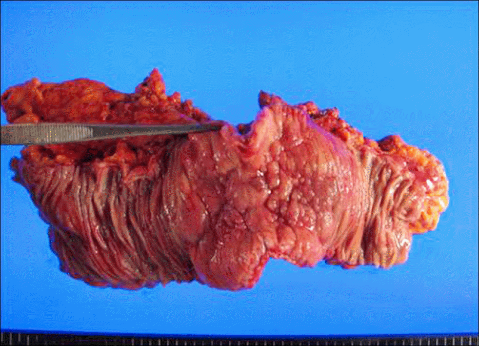
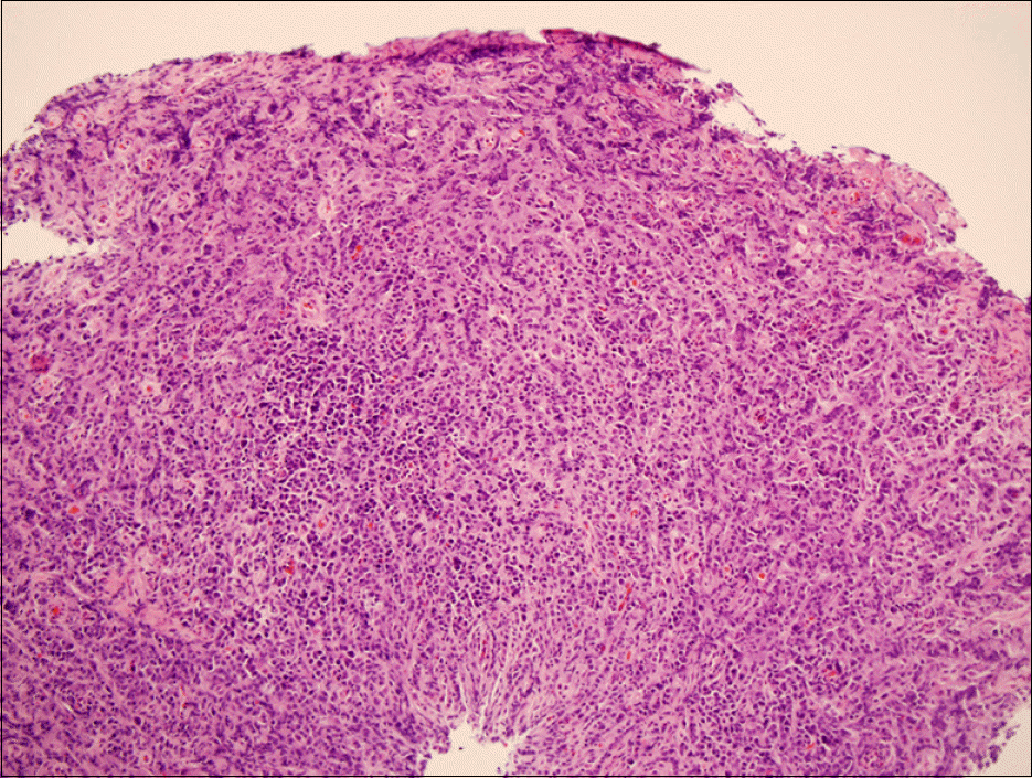
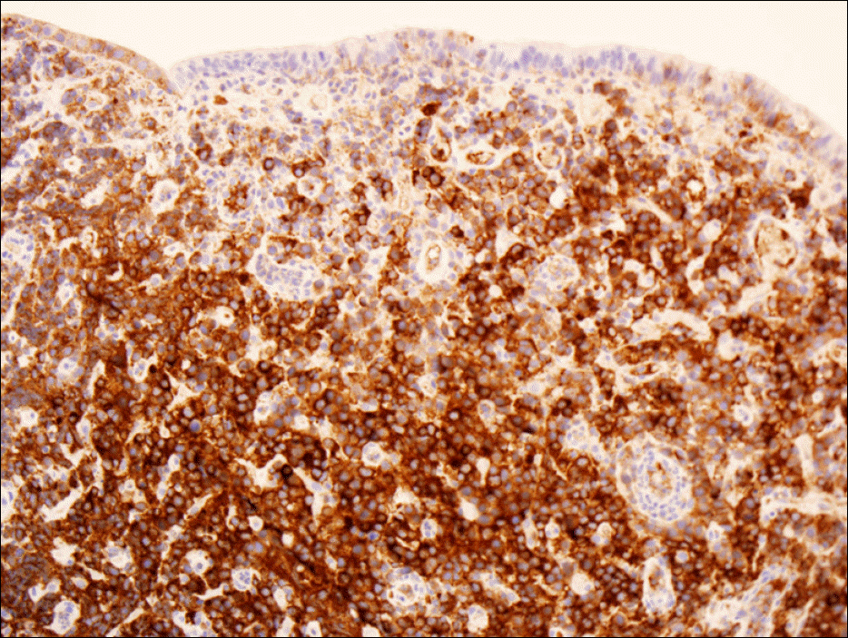
Solitary extramedullary plasmacytoma (EMP) is a plasma cell neoplasm without bone marrow involvement. EMPs are rare in the gastrointestinal (GI) tract. We report two cases of primary EMP, one in the transverse colon and the other in the stomach. In the first case, a mass on the transverse colon was found on colonoscopy. The patient underwent left hemicolectomy and has been followed-up for 3 years without recurrence postoperatively. The latter case had several masses in the stomach. The patient underwent bypass surgery and has received supportive care for 1 month. Histopathologic specimens of both the cases showed a monoclonal lambda chain EMP. Subsequent investigations included a bone marrow biopsy, serum IgA, IgG, IgM and serum protein electrophoresis, and the results were negative for multiple myeloma in both the cases. Solitary EMP in the GI tract can be mistaken for colon cancer or stomach cancer on endoscopy; therefore, a sufficient number of biopsy specimens can help diagnose solitary EMPs. Surgical resection alone or with radiation therapy in cases with positive surgical margin is currently the only treatment for solitary EMP in the GI tract. Further study is necessary to determine disease prognosis and to investigate other treatment methods.
References
1. Southerst D, Dufton J, Stern P. Multiple Myeloma presenting as sacroiliac joint pain: a case report. J Can Chiropr Assoc. 2012; 56:94–101.
2. Nolan KD, Mone MC, Nelson EW. Plasma cell neoplasms. Review of disease progression and report of a new variant. Surg Oncol. 2005; 14:85–90.
3. Alexiou C, Kau RJ, Dietzfelbinger H, et al. Extramedullary plasmacytoma: tumor occurrence and therapeutic concepts. Cancer. 1999; 85:2305–2314.
4. Weber DM. Solitary bone and extramedullary plasmacytoma. Hematology Am Soc Hematol Educ Program. 2005; 2005:373–376.

5. Kilciksiz S, Karakoyun-Celik O, Agaoglu FY, Haydaroglu A. A review for solitary plasmacytoma of bone and extramedullary plasmacytoma. Scientific World Journal. 2012; 2012:895765.
6. Dimopoulos MA, Kiamouris C, Moulopoulos LA. Solitary plasmacytoma of bone and extramedullary plasmacytoma. Hematol Oncol Clin North Am. 1999; 13:1249–1257.
7. Shin OR, Park GS, Lee YS, Jung ES, Kim SM, Kim BK. Primary extramedullary plasmacytoma of the colon: a case report. Korean J Pathol. 2001; 35:80–82.
8. Gleason TH, Hammar SP. Plasmacytoma of the colon: case report with lambda light chain, demonstrated by immunoperox-idase studies. Cancer. 1982; 50:130–133.
9. Lopes da Silva R. Extramedullary plasmacytoma of the small intestine: clinical features, diagnosis and treatment. J Dig Dis. 2012; 13:10–18.
10. Krishnamoorthy N, Bal MM, Ramadwar M, Deodhar K, Mohandas KM. A rare case of primary gastric plasmacytoma: an unforeseen surprise. J Cancer Res Ther. 2010; 6:549–551.
11. Park CH, Lee SM, Kim TO, et al. Treatment of solitary extramedullary plasmacytoma of the stomach with endoscopic submucosal dissection. Gut Liver. 2009; 3:334–337.
12. Pentimone F, Camici M, Cini G, Levorato D. Duodenal plasmacytoma. A rare primary extramedullary localization simulating a carcinoma. Acta Haematol. 1979; 61:155–160.
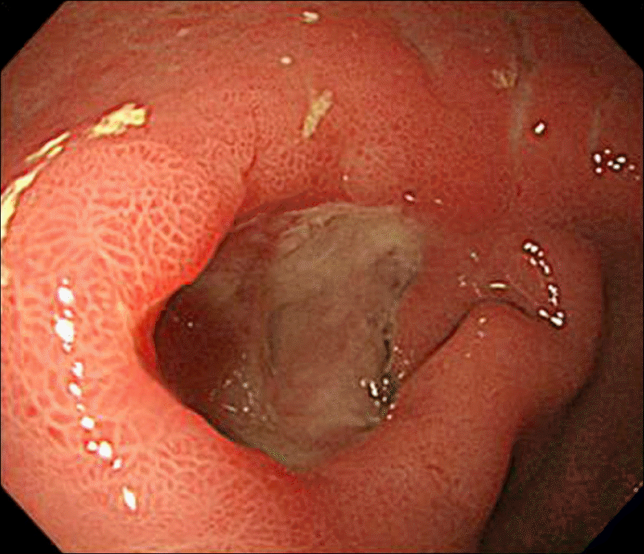
Fig. 1.
Colonoscopy showed edematous and nodular mucosal changes 45–55 cm from the anal verge, and a mass-like lesion that protruded into the lumen of the transverse colon 50 cm from the anal verge.
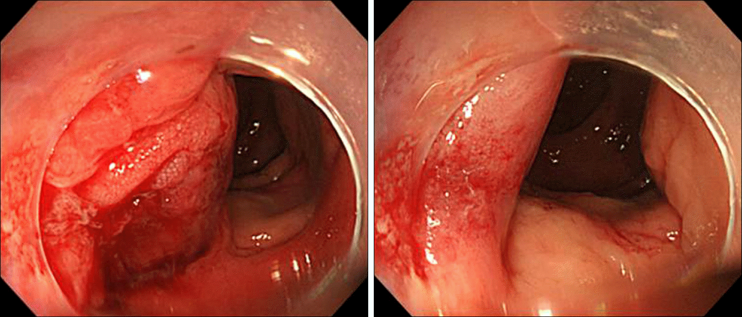
Fig. 2.
CT showed enhancing circumferential wall thickening and luminal narrowing (arrow) in the transverse colon (portal phase).
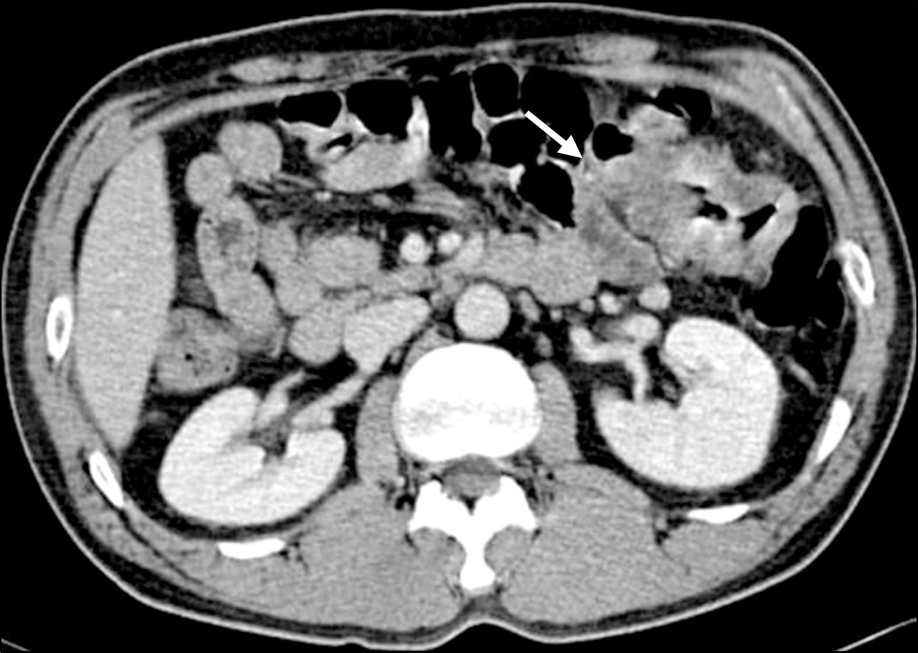
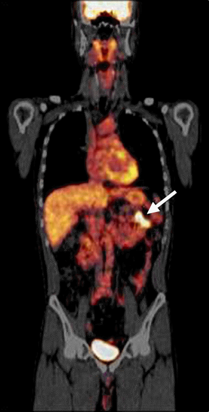
Fig. 3.
Hypermetabolism in the transverse colon (arrow) (max SUV=7.7) without distant metastasis was observed on PET-CT.
 XML Download