References
1. Choi JM, Lee C, Han YM, et al. Clinical characteristics of lower gastrointestinal cancer in crohn's disease: case series of 5 patients. Intest Res. 2013; 11:127–133.

2. Yang SK, Yun S, Kim JH, et al. Epidemiology of inflammatory bowel disease in the Songpa-Kangdong district, Seoul, Korea, 1986–2005: a KASID study. Inflamm Bowel Dis. 2008; 14:542–549.

3. Mizushima T, Ohno Y, Nakajima K, et al. Malignancy in Crohn's disease: incidence and clinical characteristics in Japan. Digestion. 2010; 81:265–270.

4. Tirkes AT, Duerinckx AJ. Adenocarcinoma of the ileum in Crohn disease. Abdom Imaging. 2005; 30:671–673.

5. Shin DW, Hahn BC, Shin JU, et al. A case of colon carcinoma in crohn's disease. Korean J Med. 2000; 59:80–84.
6. Kim JS, Cheung DY, Park SH, et al. A case of small intestinal signet ring cell carcinoma in crohn's disease. Korean J Gastroenterol. 2007; 50:51–55.
7. Warren S, Sommers SC. Cicatrizing enteritis as a pathologic en-tity; analysis of 120 cases. Am J Pathol. 1948; 24:475–501.
8. Kamiya T, Ando T, Ishiguro K, et al. Intestinal cancers occurring in patients with Crohn's disease. J Gastroenterol Hepatol. 2012; 27(Suppl 3):103–107.

9. Friedman S, Rubin PH, Bodian C, Goldstein E, Harpaz N, Present DH. Screening and surveillance colonoscopy in chronic Crohn's colitis. Gastroenterology. 2001; 120:820–826.

10. Bernstein CN, Blanchard JF, Kliewer E, Wajda A. Cancer risk in patients with inflammatory bowel disease: a population-based study. Cancer. 2001; 91:854–862.
11. Ekbom A, Helmick C, Zack M, Adami HO. Increased risk of large-bowel cancer in Crohn's disease with colonic involvement. Lancet. 1990; 336:357–359.

12. Palascak-Juif V, Bouvier AM, Cosnes J, et al. Small bowel adenocarcinoma in patients with Crohn's disease compared with small bowel adenocarcinoma de novo. Inflamm Bowel Dis. 2005; 11:828–832.

13. Solem CA, Harmsen WS, Zinsmeister AR, Loftus EV Jr. Small intestinal adenocarcinoma in Crohn's disease: a casecontrol study. Inflamm Bowel Dis. 2004; 10:32–35.
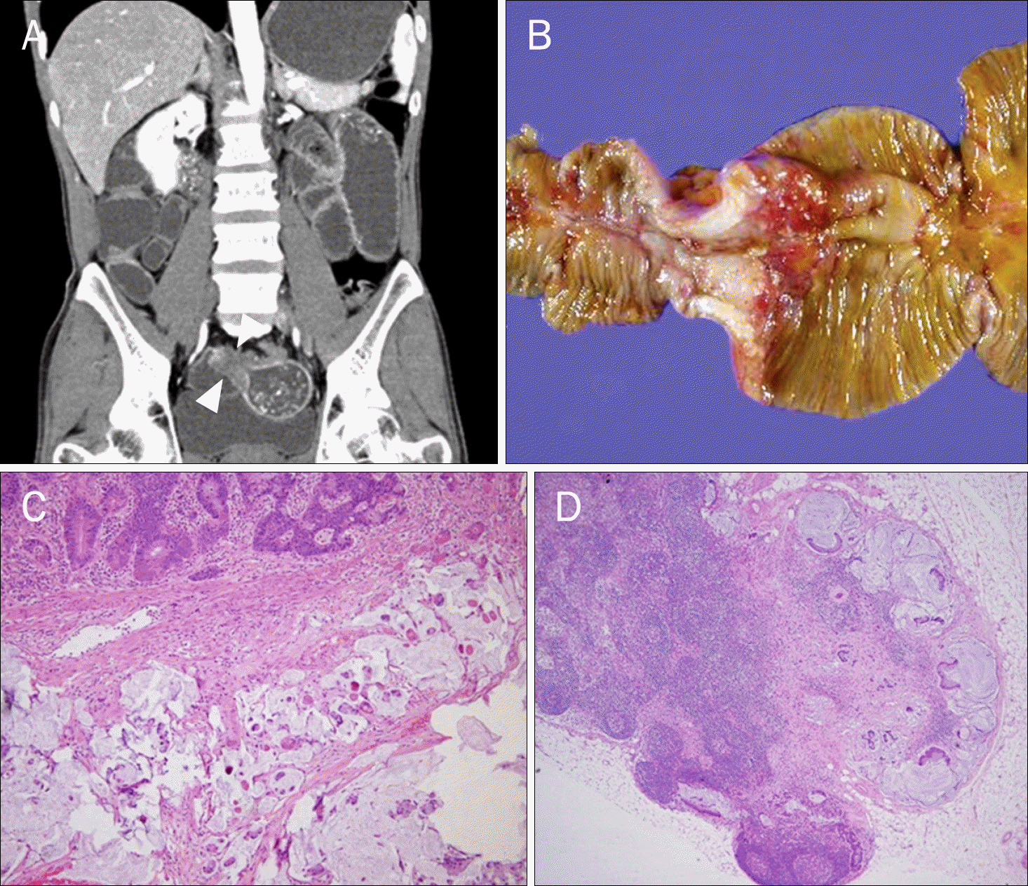
Fig. 1.
(A) Abdomen CT scan shows circumferential wall thickening (arrow-heads) of the small bowel with luminal narrowing and dilated bowel proximal to the obstruction. (B) This lesion was resected by surgery. (C) Microscopic findings show mucinous adeno-carcinoma invading the sub serosa containing signet ring cells (H&E, ×100) and (D) mesenteric lymph node metastases (H&E, ×40).

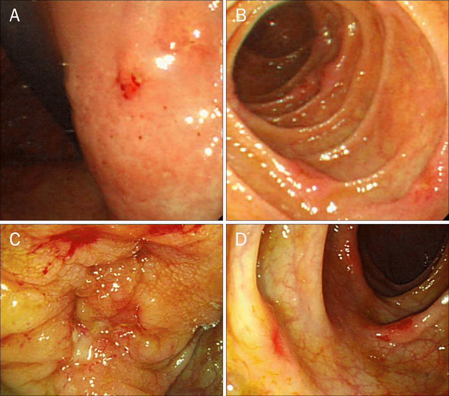
Fig. 2.
Endoscopic findings. Multiple nodular lesions with erosion are seen in the stomach (A) and duodenum (B). Colonoscopy shows hard nodular mucosa at the anastomotic site (C) and multiple variable sized ulcerations through the entire colon (D).

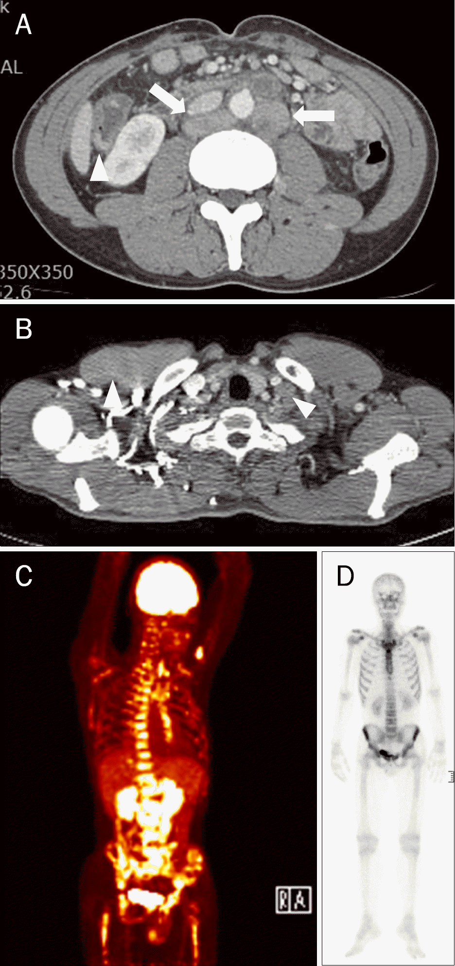
Fig. 3.
Staging workup results. (A) Abdomen CT scan shows multifocal enhancing wall thickening (arrowhead) and multiple lymph node enlargements in the retroperitoneum (arrows). (B) Chest CT showed enlarged lymph nodes in the left supraclavicular area (arrowheads), mediastinum (4B), and retrocrural area. (C) PET scan reveals hypermetabolic lymph nodes in the left supraclavicular area (maximal standardized uptake value [SUV] 5.0), mediastinum (4R: maximal SUV 5.1), retrocrural (maximal SUV 5.3), retroperitoneum (left para-aortic: maximal SUV 5.5), and both common iliac area. Additionally, irregular, partial focal hypermetabolism in marrow space is observed. (D) Bone scan shows diffuse increased uptake in whole axial and both proximal appendicular skeletons suggesting early bone marrow involvement.


 PDF
PDF ePub
ePub Citation
 Print
Print


 XML Download