References
1. Sato T, Akaike J, Toyota J, Karino Y, Ohmura T. Clinicopathological features and treatment of ectopic varices with portal hypertension. Int J Hepatol. 2011; 2011:960720.

2. Lee JY, Song SY, Kim J, et al. Percutaneous transsplenic embolization of jejunal varices in a patient with liver cirrhosis: a case report. Abdom Imaging. 2013; 38:52–55.

3. Sasamoto A, Kamiya J, Nimura Y, Nagino M. Successful embolization therapy for bleeding from jejunal varices after chol-edochojejunostomy: report of a case. Surg Today. 2010; 40:788–791.

4. Boku M, Sugimoto K, Nakamura T, Kita Y, Zamora CA, Sugimura K. Percutaneous transhepatic obliteration for bleeding esophagojejunal varices after total gastrectomy and esophagojejunostomy. Cardiovasc Intervent Radiol. 2006; 29:1152–1155.

5. Sato T, Yasui O, Kurokawa T, Hashimoto M, Asanuma Y, Koyama K. Jejunal varix with extrahepatic portal obstruction treated by embolization using interventional radiology: report of a case. Surg Today. 2003; 33:131–134.

6. Saeki Y, Ide K, Kakizawa H, Ishikawa M, Tashiro H, Ohdan H. Controlling the bleeding of jejunal varices formed at the site of choledochojejunostomy: report of 2 cases and a review of the literature. Surg Today. 2013; 43:550–555.

7. Shussman N, Lalazar G, Bloom AI. Isolated jejunal varices: a cause of occult gastrointestinal hemorrhage in a cirrhotic patient with mild portal hypertension. Clin Gastroenterol Hepatol. 2012; 10:A32.

8. Gubler C, Glenck M, Pfammatter T, Bauerfeind P. Successful treatment of anastomotic jejunal varices with N-butyl-2-cyanoa-crylate (Histoacryl): single-center experience. Endoscopy. 2012; 44:776–779.

Go to : 
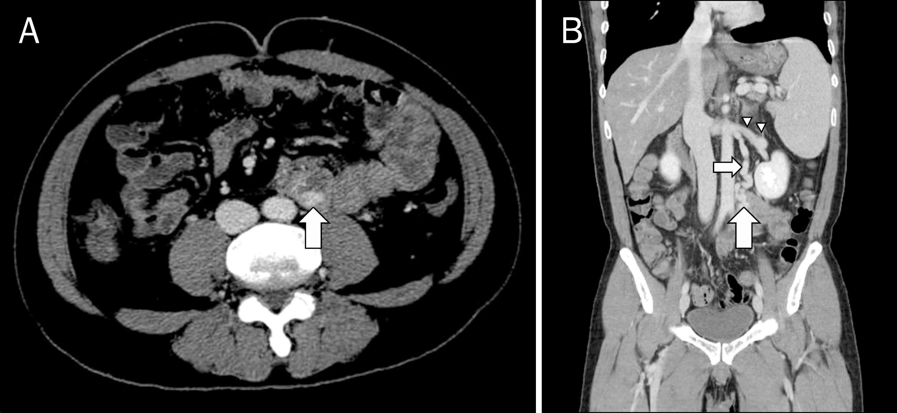
 | Fig. 1.(A) Axial CT image demonstrates an enhancing dilated vein in the intestinal wall (arrow). (B) Coronal image shows the renal vein (arrow-heads), gonadal vein (small arrow), and jejunal varix (large arrow) which are all connected. |
 | Fig. 2.Balloon-occluded retrograde transvenous obliteration of a jejunal varix. (A) Digital subtraction angiography obtained after inflation of the occlusion balloon and retrograde injection of contrast agent demonstrates that the varix and its connected mesenteric veins are draining into the superior mesenteric vein (arrowheads) and inferior mesenteric vein (arrows). (B) A spot image shows the mixture of sclerosing agent and contrast agent filling the varix. |


 PDF
PDF ePub
ePub Citation
 Print
Print


 XML Download