

 PDF
PDF ePub
ePub Citation
 Print
Print


Abstract
Background/Aims
The polyp detection rate (PDR) has been suggested as a surrogate for adenoma detection rate (ADR). The purpose of this study was to determine the level of agreement between PDR and ADR in the proximal and distal colon.
Methods
A total of 1,937 consecutive, asymptomatic individuals aged 40 years and older who underwent colonoscopies at six academic teaching hospitals in Korea were included in this study. PDR and ADR were calculated for each colonic segment. PDR was compared with ADR in the proximal and distal colon.
Results
During 1,937 colonoscopies, 1,862 polyps were removed; 1,421 (76%) were adenomas. The PDR and ADR in the proximal colon was 25.8% and 22.8%, respectively (kappa value=0.917, p=0.26), and that in the distal colon was 28.9% and 22.2%, respectively (p<0.001). There was a strong correlation between PDR and ADR in the proximal colon, but diverged in sigmoid colon and rectum.
References
1. Zauber AG, Winawer SJ, O' Brien MJ, et al. Colonoscopic polypectomy and long-term prevention of colorectal-cancer deaths. N Engl J Med. 2012; 366:687–696.

2. Atkin W, Rogers P, Cardwell C, et al. Wide variation in adenoma detection rates at screening flexible sigmoidoscopy. Gastroenterology. 2004; 126:1247–1256.
3. Rex DK. Colonoscopic withdrawal technique is associated with adenoma miss rates. Gastrointest Endosc. 2000; 51:33–36.
4. Rex DK, Cutler CS, Lemmel GT, et al. Colonoscopic miss rates of adenomas determined by back-to-back colonoscopies. Gastroenterology. 1997; 112:24–28.
5. Sanchez W, Harewood GC, Petersen BT. Evaluation of polyp detection in relation to procedure time of screening or surveillance colonoscopy. Am J Gastroenterol. 2004; 99:1941–1945.
6. Kaminski MF, Regula J, Kraszewska E, et al. Quality indicators for colonoscopy and the risk of interval cancer. N Engl J Med. 2010; 362:1795–1803.
7. Rex DK, Bond JH, Winawer S, et al. U.S. MultiSociety Task Force on Colorectal Cancer. Quality in the technical performance of colonoscopy and the continuous quality improvement process for colonoscopy: recommendations of the U.S. MultiSociety Task Force on Colorectal Cancer. Am J Gastroenterol. 2002; 97:1296–1308.
8. Lee SH, Park DI, Sung JM, et al. Usefulness of polyp detection rate as a quality indicator in colonoscopy. Intest Res. 2011; 9:105–111.
9. Winawer SJ, Zauber AG, Ho MN, et al. Prevention of colorectal cancer by colonoscopic polypectomy. The National Polyp Study Workgroup. N Engl J Med. 1993; 329:1977–1981.
10. Brenner H, Chang-Claude J, Seiler CM, Rickert A, Hoffmeister M. Protection from colorectal cancer after colonoscopy: a population-based, case-control study. Ann Intern Med. 2011; 154:22–30.
11. Citarda F, Tomaselli G, Capocaccia R, Barcherini S, Crespi M. Italian Multicentre Study Group. Efficacy in standard clinical practice of colonoscopic polypectomy in reducing colorectal cancer incidence. Gut. 2001; 48:812–815.
12. Boroff ES, Gurudu SR, Hentz JG, Leighton JA, Ramirez FC. Polyp and adenoma detection rates in the proximal and distal colon. Am J Gastroenterol. 2013; 108:993–999.
13. Rex DK, Lehman GA, Ulbright TM, et al. Colonic neoplasia in asymptomatic persons with negative fecal occult blood tests: influence of age, gender, and family history. Am J Gastroenterol. 1993; 88:825–831.
14. Johnson DA, Gurney MS, Volpe RJ, et al. A prospective study of the prevalence of colonic neoplasms in asymptomatic patients with an age-related risk. Am J Gastroenterol. 1990; 85:969–974.
15. Rogge JD, Elmore MF, Mahoney SJ, et al. Low-cost, office-based, screening colonoscopy. Am J Gastroenterol. 1994; 89:1775–1780.
16. Lieberman DA, Weiss DG, Bond JH, Ahnen DJ, Garewal H, Chejfec G. Use of colonoscopy to screen asymptomatic adults for colorectal cancer. Veterans Affairs Cooperative Study Group 380. N Engl J Med. 2000; 343:162–168.
17. Francis DL, Rodriguez-Correa DT, Buchner A, Harewood GC, Wallace M. Application of a conversion factor to estimate the adenoma detection rate from the polyp detection rate. Gastrointest Endosc. 2011; 73:493–497.
18. Barclay RL, Vicari JJ, Doughty AS, Johanson JF, Greenlaw RL. Colonoscopic withdrawal times and adenoma detection during screening colonoscopy. N Engl J Med. 2006; 355:2533–2541.
19. Rex DK. Maximizing detection of adenomas and cancers during colonoscopy. Am J Gastroenterol. 2006; 101:2866–2877.
20. Choi SW, Park HS, Lee JS, Hwang SY, Kwak SD, Choi SH. Efficacy of hood-cap assisted colonoscopy; comparison with conventional colonoscopy. Intest Res. 2012; 10:280–288.
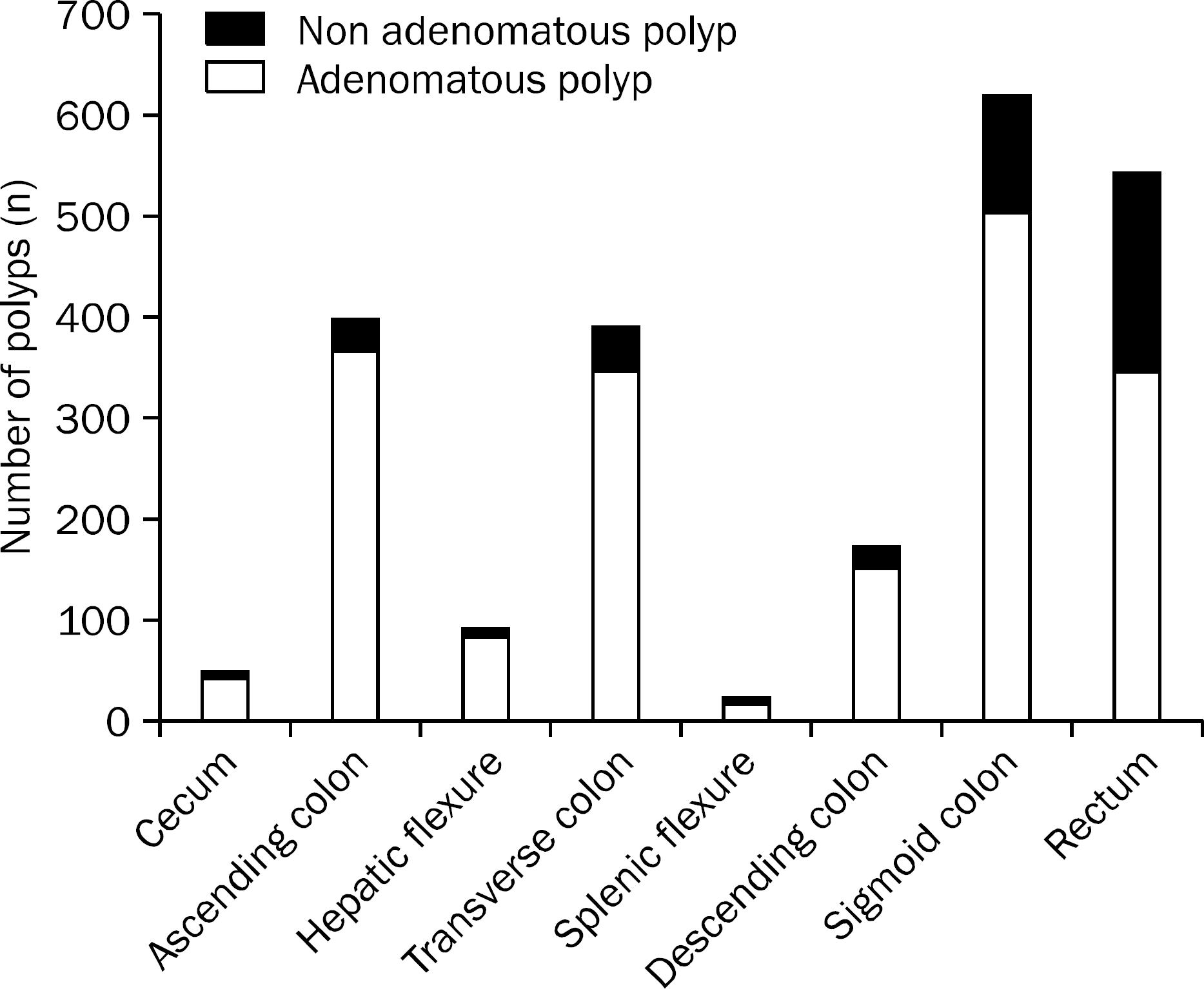
Fig. 2.
Polyp detection rate (PDR) and adenoma detection rate (ADR) by colon segment. The PDR and ADR in the proximal colon was 25.8% and 22.8%, respectively (kappa value [K]=0.917, p=0.26), and that in the distal colon was 28.9% and 22.2%, respectively (p<0.001).
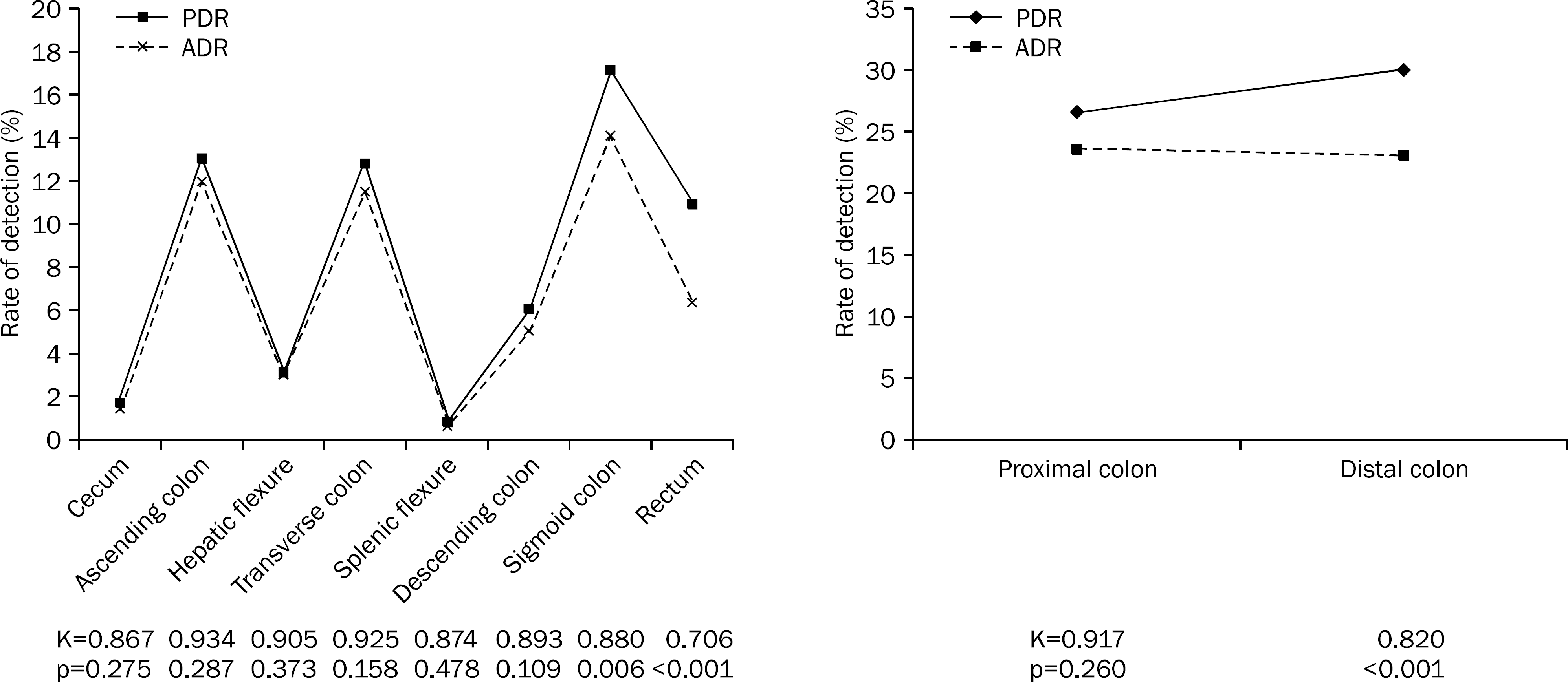
Fig. 3.
(A) Polyp detection rate (PDR) and adenoma detection rate (ADR) by colon segment in men. The PDR and ADR in the proximal colon was 31.6% and 28.3%, respectively (kappa value [K]=0.916, p=0.077), and that in the distal colon was 34.4% and 26.4%, respectively (p<0.001).(B) PDR and ADR by colon segment in women. The PDR and ADR in the proximal colon was 18.5% and 16.1%, respectively (kappa value=0.915, p=0.161), and that in the distal colon was 22.2% and 16.9%, respectively (p=0.004).
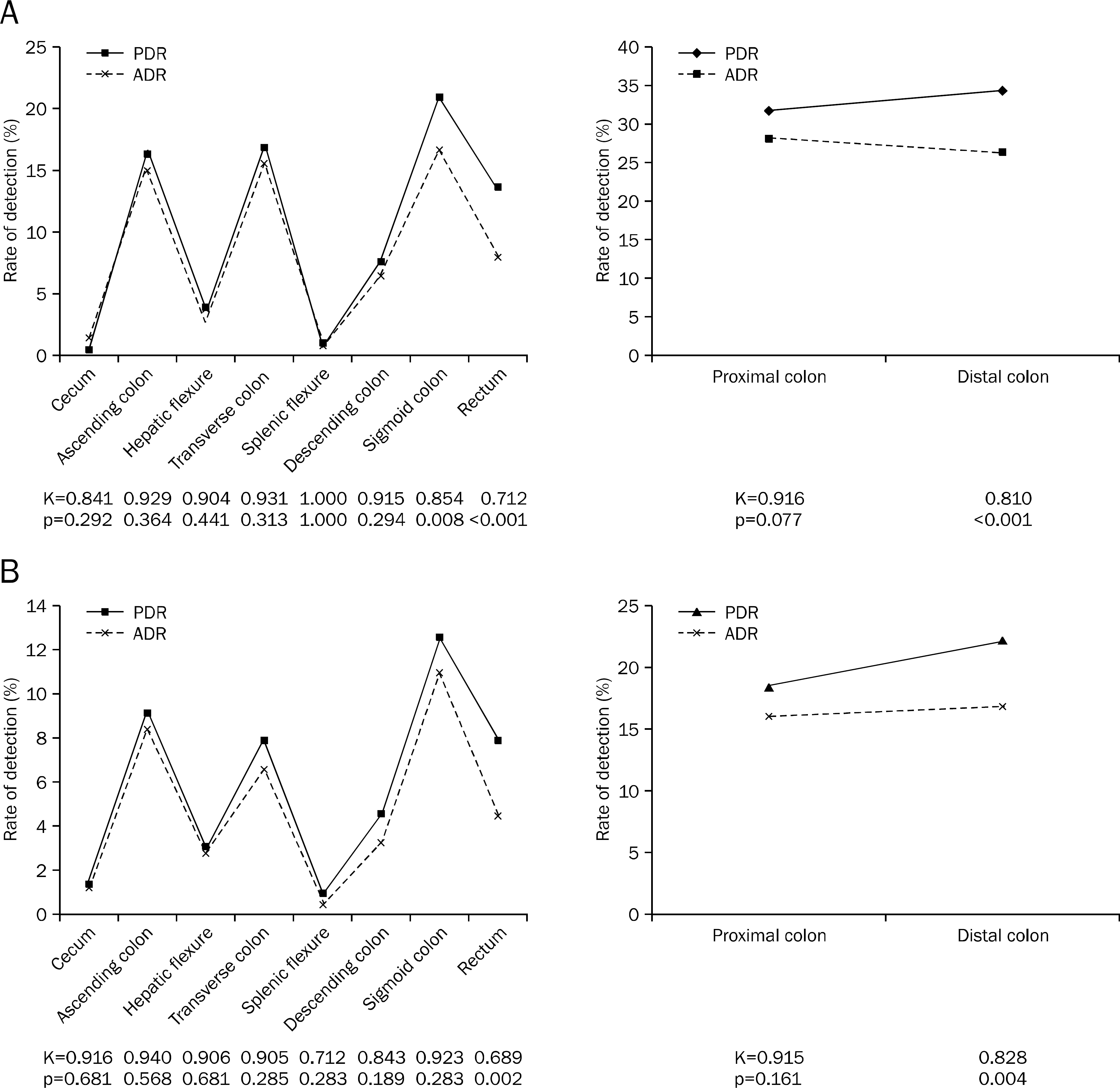
Table 1.
Demographic Features of Patients
| Factor | Value |
|---|---|
| Sex | |
| Male | 1,079 (55.7) |
| Female | 858 (44.3) |
| Age (yr) | |
| Mean | 55.4 (40–84) |
| 40–49 | 251 (13.0) |
| 50–59 | 962 (49.7) |
| 60–69 | 537 (27.7) |
| 70–79 | 179 (9.2) |
| 80–84 | 8 (0.4) |
Table 2.
Endoscopic Features of Patients
Table 3.
Proportion of Adenomas/Polyps by Size and Morphology
 XML Download