

 PDF
PDF ePub
ePub Citation
 Print
Print


References
1. Rashid F, Thangarajah T, Mulvey D, Larvin M, Iftikhar SY. A review article on gastric volvulus: a challenge to diagnosis and management. Int J Surg. 2010; 8:18–24.

2. Wasselle JA, Norman J. Acute gastric volvulus: pathogenesis, diagnosis, and treatment. Am J Gastroenterol. 1993; 88:1780–1784.
3. Shivanand G, Seema S, Srivastava DN, et al. Gastric volvulus: acute and chronic presentation. Clin Imaging. 2003; 27:265–268.
4. Chau B, Dufel S. Gastric volvulus. Emerg Med J. 2007; 24:446–447.
Fig. 1.
Endoscopic findings. (A) The large amount of gastric juice and food was seen in the stomach. Endoscopic approach from the fundus to lower body was impossible. White arrow is pointing direction to the lower body. (B) The entry of hernia sac (black arrow) was seen at the fundus.
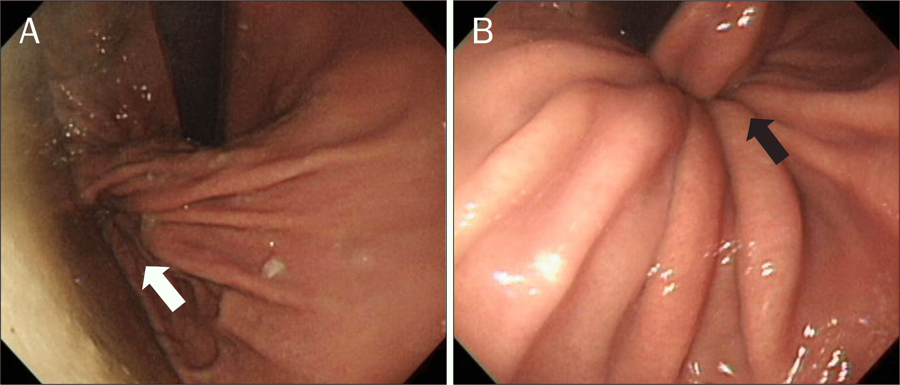
Fig. 2.
Computed tomography findings. (A) It showed marked distension of the upper body and fundus of the stomach and air-fluid level. Lower part of the stomach was collapsed and twisted (black arrow). (B) The hernia sac was seen in the thoracic cavity (white arrow).

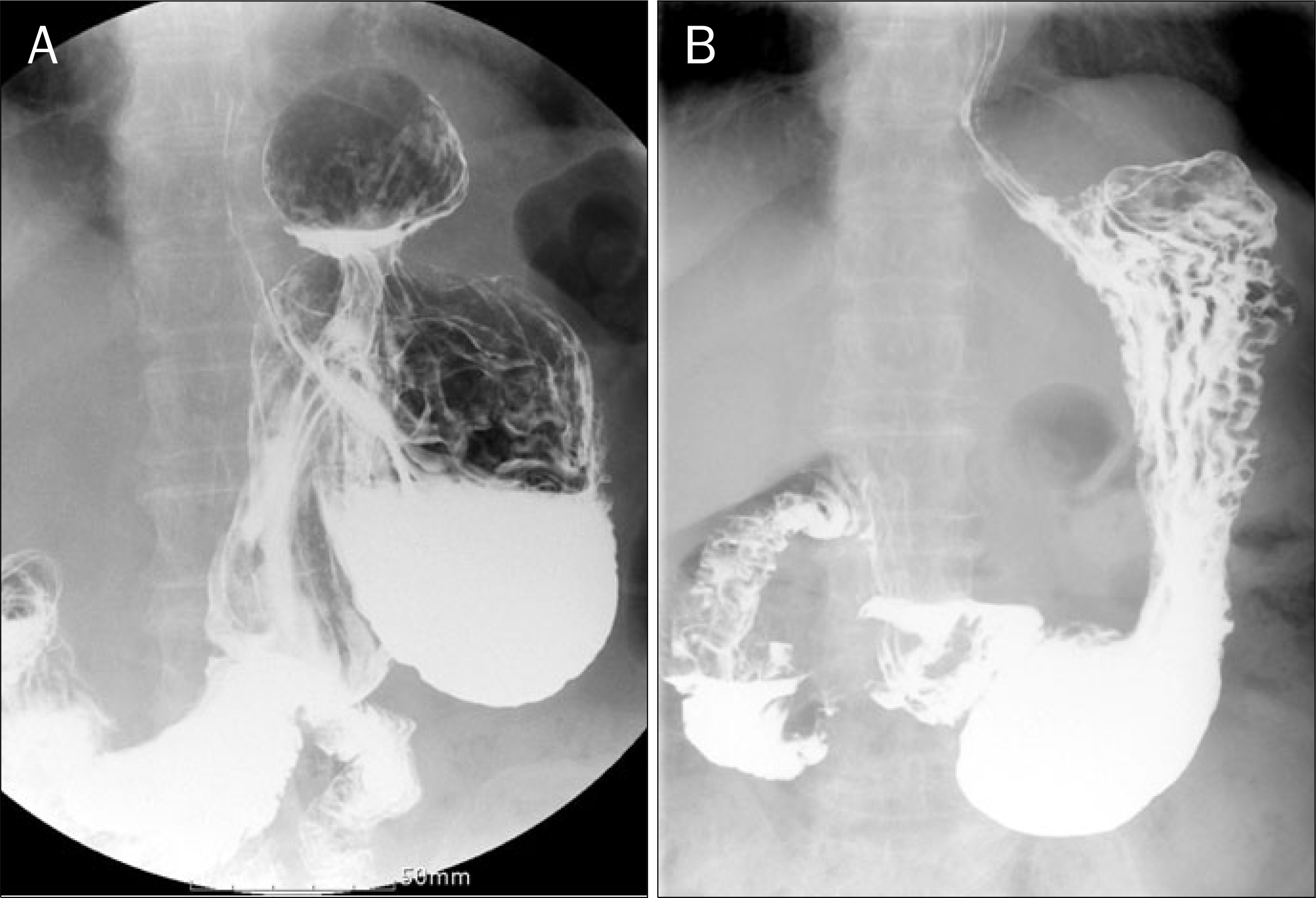
Fig. 3.
(A) Preoperative upper gastrointestinal series. It showed hernia sac, dilated upper body and fundus with underflow of barium, and twisted and collapsed lower body and antrum. (B) Follow-up of postoperative upper gastrointestinal series. It showed normal anatomical structure of stomach without herniated sac.
 XML Download