Abstract
Background/Aims
Metabolic syndrome is a well-known risk factor for atherosclerosis. Non-alcoholic fatty liver disease (NAFLD) has features of metabolic syndromes. This study aimed to investigate the association between NAFLD and atherosclerosis.
Methods
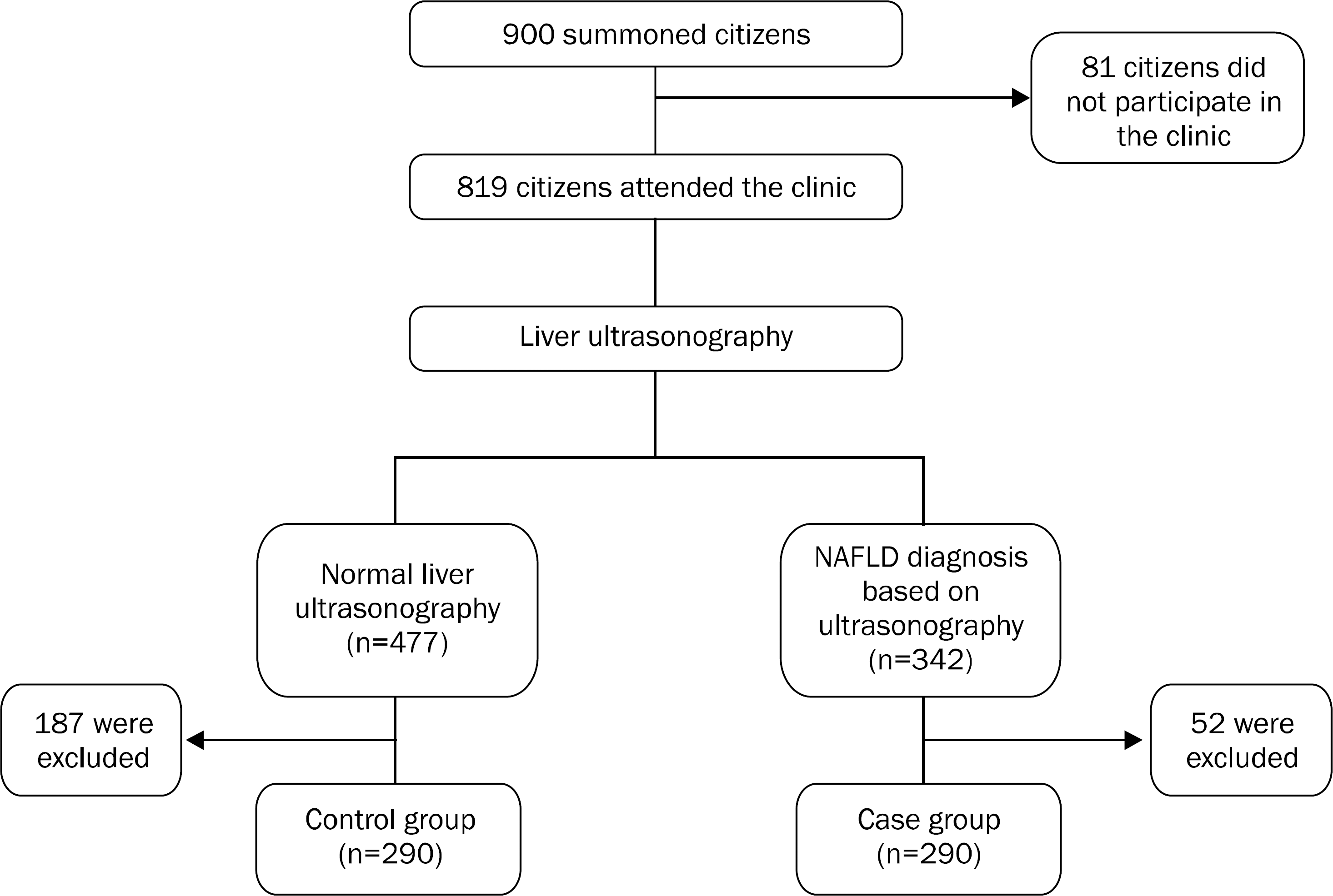
In a population-based study in southern Iran, asymptomatic adult inhabitants aged more than 20 years were selected through cluster random sampling, and were screened for the presence of fatty liver and common carotid intima-media thickness (CIMT), with abdominal and cervical ultrasonography, respectively. Those with fatty liver were compared to the same number of individuals without fatty liver.
Results
Two hundred and ninety individuals were found to have fatty change on abdominal ultrasonography, and were labeled NAFLD. Compared to normal individuals, NAFLD patients had significantly higher prevalence of increased CIMT (OR, 1.66; p<0.001). Those with hypertension (HTN), diabetes mellitus (DM), higher waist circumference (WC) and older ages had significantly higher prevalence of thick CIMT. Through adjusting the effects of different variables, we indicated that NAFLD could be an independent risk factor for thick common carotid intima-media (OR, 1.90; 95% CI, 1.17–3.09; p=0.009). It was also shown that age could be another independent risk factor for thick CIMT.
Go to : 
References
2. Levene AP, Goldin RD. The epidemiology, pathogenesis and histopathology of fatty liver disease. Histopathology. 2012; 61:141–152.

3. Vernon G, Baranova A, Younossi ZM. Systematic review: the epidemiology and natural history of non-alcoholic fatty liver disease and non-alcoholic steatohepatitis in adults. Aliment Pharmacol Ther. 2011; 34:274–285.

4. Ahmed MH, Abu EO, Byrne CD. Non-alcoholic fatty liver disease (NAFLD): new challenge for general practitioners and important burden for health authorities? Prim Care Diabetes. 2010; 4:129–137.

5. Lankarani KB, Ghaffarpasand F, Mahmoodi M, et al. Non alcoholic fatty liver disease in southern Iran: a population based study. Hepat Mon. 2013; 13:e9248.

6. Lee SY, Kim SK, Kwon CI, et al. Clinical characteristics of health screen examinees with nonalcoholic fatty liver and normal liver function test. Korean J Gastroenterol. 2008; 52:161–170.
7. Margariti E, Deutsch M, Manolakopoulos S, Papatheodoridis GV. Non-alcoholic fatty liver disease may develop in individuals with normal body mass index. Annals of Gastroenterology. 2012; 25:45–51.
8. Sookoian S, Pirola CJ. Non-alcoholic fatty liver disease is strongly associated with carotid atherosclerosis: a systematic review. J Hepatol. 2008; 49:600–607.

9. Basevi V, Di Mario S, Morciano C, Nonino F, Magrini N. Comment on: American Diabetes Association. Standards of medical care in diabetes–2011. Diabetes Care. 2011; 34(Suppl. 1):S11–S61.
10. Lenfant C, Chobanian AV, Jones DW, Roccella EJ. Joint National Committee on the Prevention, Detection, Evaluation, and Treatment of High Blood Pressure. Seventh report of the Joint National Committee on the Prevention, Detection, Evaluation, and Treatment of High Blood Pressure (JNC 7): resetting the hypertension sails. Hypertension. 2003; 41:1178–1179.
11. Grundy SM, Cleeman JI, Daniels SR, et al. American Heart Association; National Heart, Lung, and Blood Institute. Diagnosis and management of the metabolic syndrome: an American Heart Association/National Heart, Lung, and Blood Institute Scientific Statement. Circulation. 2005; 112:2735–2752.
12. Saadeh S, Younossi ZM, Remer EM, et al. The utility of radiological imaging in nonalcoholic fatty liver disease. Gastroenterology. 2002; 123:745–750.

13. Dougherty RS, Brant WE. Vascular ultrasound. Brant WE, Helms CA, editors. Fundamentals of diagnostic radiology. 3rd ed.Philadelphia: Lippincott, Williams & Wilkins;2007. p. 1027.
14. Yki-Järvinen H. Nutritional modulation of nonalcoholic fatty liver disease and insulin resistance: human data. Curr Opin Clin Nutr Metab Care. 2010; 13:709–714.

15. Kotronen A, Westerbacka J, Bergholm R, Pietiläinen KH, Yki-Järvinen H. Liver fat in the metabolic syndrome. J Clin Endocrinol Metab. 2007; 92:3490–3497.

16. Musso G, Gambino R, Bo S, et al. Should nonalcoholic fatty liver disease be included in the definition of metabolic syndrome? A cross-sectional comparison with Adult Treatment Panel III criteria in nonobese nondiabetic subjects. Diabetes Care. 2008; 31:562–568.

17. Targher G, Day CP, Bonora E. Risk of cardiovascular disease in patients with nonalcoholic fatty liver disease. N Engl J Med. 2010; 363:1341–1350.

18. Targher G, Bertolini L, Rodella S, et al. Nonalcoholic fatty liver disease is independently associated with an increased incidence of cardiovascular events in type 2 diabetic patients. Diabetes Care. 2007; 30:2119–2121.

19. Maurantonio M, Ballestri S, Odoardi MR, Lonardo A, Loria P. Treatment of atherogenic liver based on the pathogenesis of nonalcoholic fatty liver disease: a novel approach to reduce cardiovascular risk? Arch Med Res. 2011; 42:337–353.

20. Loria P, Adinolfi LE, Bellentani S, et al. NAFLD Expert Committee of the Associazione Italiana per lo studio del Fegato. Practice guidelines for the diagnosis and management of nonalcoholic fatty liver disease. A decalogue from the Italian Association for the Study of the Liver (AISF) Expert Committee. Dig Liver Dis. 2010; 42:272–282.
21. Targher G, Bertolini L, Padovani R, et al. Relations between carotid artery wall thickness and liver histology in subjects with non-alcoholic fatty liver disease. Diabetes Care. 2006; 29:1325–1330.

22. Karakurt F, Carlioglu A, Koktener A, et al. Relationship between cerebral arterial pulsatility and carotid intima media thickness in diabetic and non-diabetic patients with non-alcoholic fatty liver disease. J Endocrinol Invest. 2009; 32:63–68.

23. Ying I, Saposnik G, Vermeulen MJ, Leung A, Ray JG. Nonalcoholic fatty liver disease and acute ischemic stroke. Epidemiology. 2011; 22:129–130.

24. Targher G, Day CP, Bonora E. Risk of cardiovascular disease in patients with nonalcoholic fatty liver disease. N Engl J Med. 2010; 363:1341–1350.

25. Thakur ML, Sharma S, Kumar A, et al. Nonalcoholic fatty liver disease is associated with subclinical atherosclerosis independent of obesity and metabolic syndrome in Asian Indians. Atherosclerosis. 2012; 223:507–511.

26. Assy N, Djibre A, Farah R, Grosovski M, Marmor A. Presence of coronary plaques in patients with nonalcoholic fatty liver disease. Radiology. 2010; 254:393–400.

28. Manousou P, Kalambokis G, Grillo F, et al. Serum ferritin is a discriminant marker for both fibrosis and inflammation in histologically proven non-alcoholic fatty liver disease patients. Liver Int. 2011; 31:730–739.

29. Marchesini G, Bugianesi E, Forlani G, et al. Nonalcoholic fatty liver, steatohepatitis, and the metabolic syndrome. Hepatology. 2003; 37:917–923.

30. Zelber-Sagi S, Nitzan-Kaluski D, Halpern Z, Oren R. NAFLD and hyperinsulinemia are major determinants of serum ferritin levels. J Hepatol. 2007; 46:700–707.

31. Pang JH, Jiang MJ, Chen YL, et al. Increased ferritin gene expression in atherosclerotic lesions. J Clin Invest. 1996; 97:2204–2212.

32. Brea A, Mosquera D, Martín E, Arizti A, Cordero JL, Ros E. Nonalcoholic fatty liver disease is associated with carotid atherosclerosis: a case-control study. Arterioscler Thromb Vasc Biol. 2005; 25:1045–1050.
33. Fracanzani AL, Burdick L, Raselli S, et al. Carotid artery in-tima-media thickness in nonalcoholic fatty liver disease. Am J Med. 2008; 121:72–78.

34. Kim HC, Kim DJ, Huh KB. Association between nonalcoholic fatty liver disease and carotid intima-media thickness according to the presence of metabolic syndrome. Atherosclerosis. 2009; 204:521–525.

35. Nambi V, Chambless L, He M, et al. Common carotid artery in-tima-media thickness is as good as carotid intima-media thickness of all carotid artery segments in improving prediction of coronary heart disease risk in the Atherosclerosis Risk in Communities (ARIC) study. Eur Heart J. 2012; 33:183–190.

Go to : 
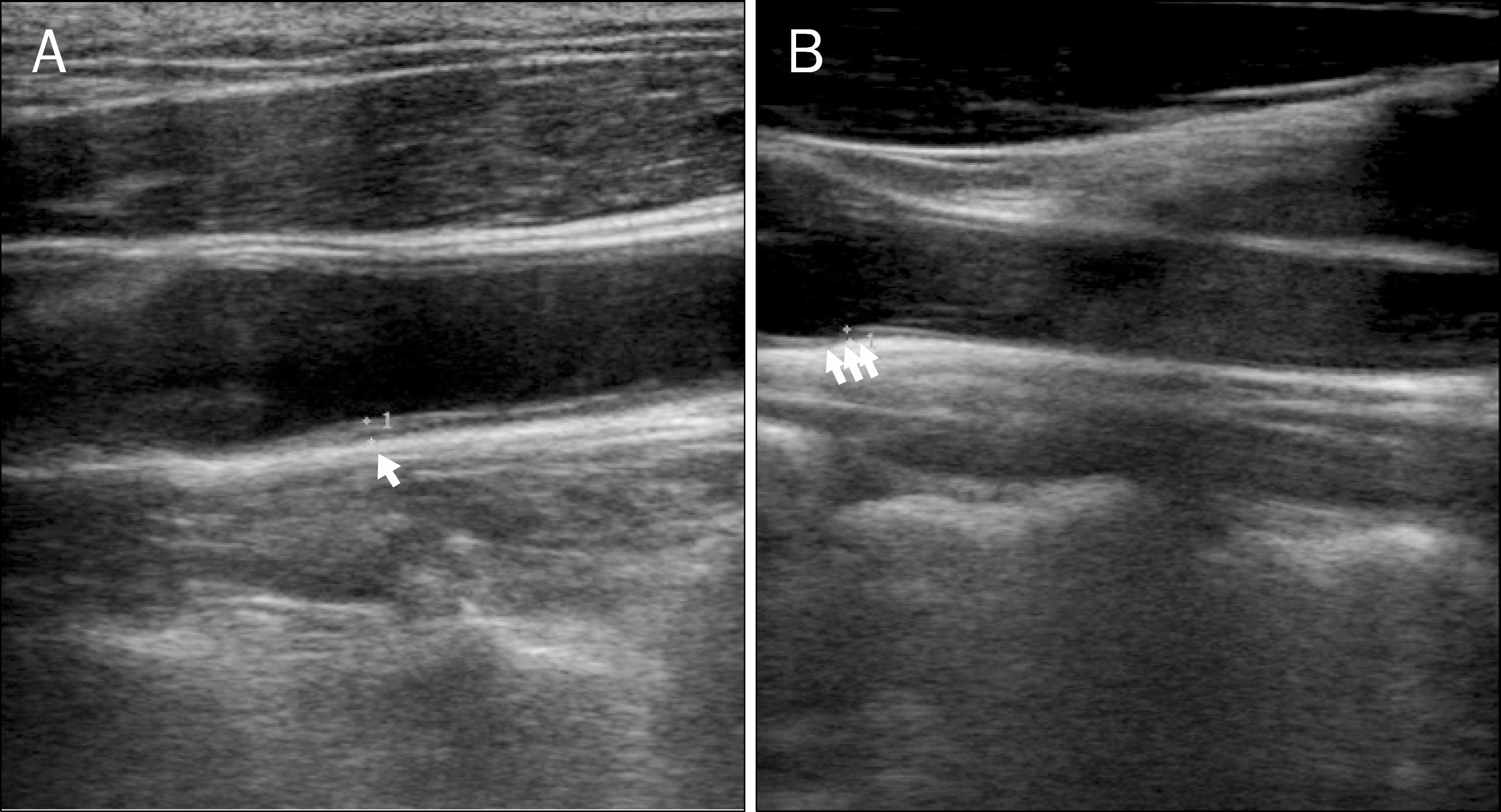
 | Fig. 2.Longitudinal ultrasonographic image of the common carotid artery. Longitudinal ultrasonographic image of the common carotid artery, showing the carotid intima-media thickness in the posterior wall (diameter: 11 mm [A], 7 mm [B]). |
Table 1.
Clinical, Anthropometric and Laboratory Findings of Patients with NAFLD and Control Group (Ultrasonographic Normal Liver Group)
Table 2.
Comparison between CIMT and Metabolic Syndrome- related Factors
For the comparison between those with thick CIMT and subjects with normal CIMT, student's t-test were used for quantitative variables, and the chi-square test was used to determine the statistical significance of differences in qualitative variables. p-value less than 0.05 is considered statistically significant.
Table 3.
Multivariate Analysis for the Risk Factors of CIMT ≥0.8 mma
| Variable | p-value | OR (CIMT ≥0.8 mm/<0.8 mm) | 95% CI |
|---|---|---|---|
| NAFLD | |||
| No | 1 | − | |
| Yes | 0.009 | 1.91 | 1.17–3.10 |
| Hypertension | |||
| No | 1 | − | |
| Yes | 0.238 | 1.34 | 0.82–2.19 |
| Diabetes mellitus | |||
| No | 1 | − | |
| Yes | 0.611 | 1.14 | 0.68–1.89 |
| Waist circumferenceb | |||
| Normal | 1 | − | |
| High | 0.054 | 0.62 | 0.38–1.01 |
| Age (yr) | |||
| 18–29 | 1 | − | |
| 30–39 | 0.166 | 0.27 | 0.04–1.710 |
| 40–49 | 0.048 | 3.50 | 1.01–12.18 |
| 50–59 < | <0.001 | 10.83 | 3.07–38.20 |
| ≥60 < | <0.001 | 22.76 | 6.08–85.21 |


 PDF
PDF ePub
ePub Citation
 Print
Print


 XML Download