Spontaneous Isolated Superior Mesenteric Artery Dissection Mimicking Superior Mesenteric Artery Syndrome
Wook Hyun Lee, Chul-Hyun Lim, Sang Woo Kim
Department of Internal Medicine, The Catholic University of Korea College of Medicine, Seoul, Korea
Correspondence to: Chul-Hyun Lim, Department of Internal Medicine, The Catholic University of Korea, Seoul St. Mary's Hospital, 222 Banpo-daero, Seocho-gu, Seoul 137-701, Korea. Tel: +82-2-2258-2083, Fax: +82-2-2258-2055, E-mail: diluck@catholic.ac.kr
This is an Open Access article distributed under the terms of the Creative Commons Attribution Non-Commercial License (http://creativecommons.org/licenses/by-nc/3.0/) which permits unrestricted non-commercial use, distribution, and reproduction in any medium, provided the original work is properly cited.
This article has been cited by other articles in ScienceCentral.
References
1. Katsura M, Mototake H, Takara H, Matsushima K. Management of spontaneous isolated dissection of the superior mesenteric artery: case report and literature review. World J Emerg Surg. 2011; 6:16.
2. Javerliat I, Becquemin JP, d' Audiffret A. Spontaneous isolated dissection of the superior mesenteric artery. Eur J Vasc Endovasc Surg. 2003; 25:180–184.
3. Min SI, Yoon KC, Min SK, et al. Current strategy for the treatment of symptomatic spontaneous isolated dissection of superior mesenteric artery. J Vasc Surg. 2011; 54:461–466.
Fig. 1.
Abdominal CT scan reveals marked distension of stomach and duodenum (A, B), and narrowing of proximal jejunum (C). (D) An 8×6 mm-sized thrombus is seen within the proximal superior mesenteric artery.
Fig. 2.
(A) Angiography shows moderate and eccentric stenosis of the proximal superior mesenteric artery. (B) An 8 Fr guiding catheter was inserted into arterial sheath over the 0.035 Fr guidewire and its tip was placed within the proximal portion of the superior mesenteric artery.(C, D) The stenotic site was dilated and an 8×40 mm self expandable stent (Zilver; Cook Medical, Bloomington, IN, USA) was inserted which was expanded wtih 6×20 mm balloon (Foxcross; Abbott Vascular, Abbott Park, IL, USA). (E) Follow-up renal arteriography reveals well positioned stent with patent superior mesenteric artery lumen.
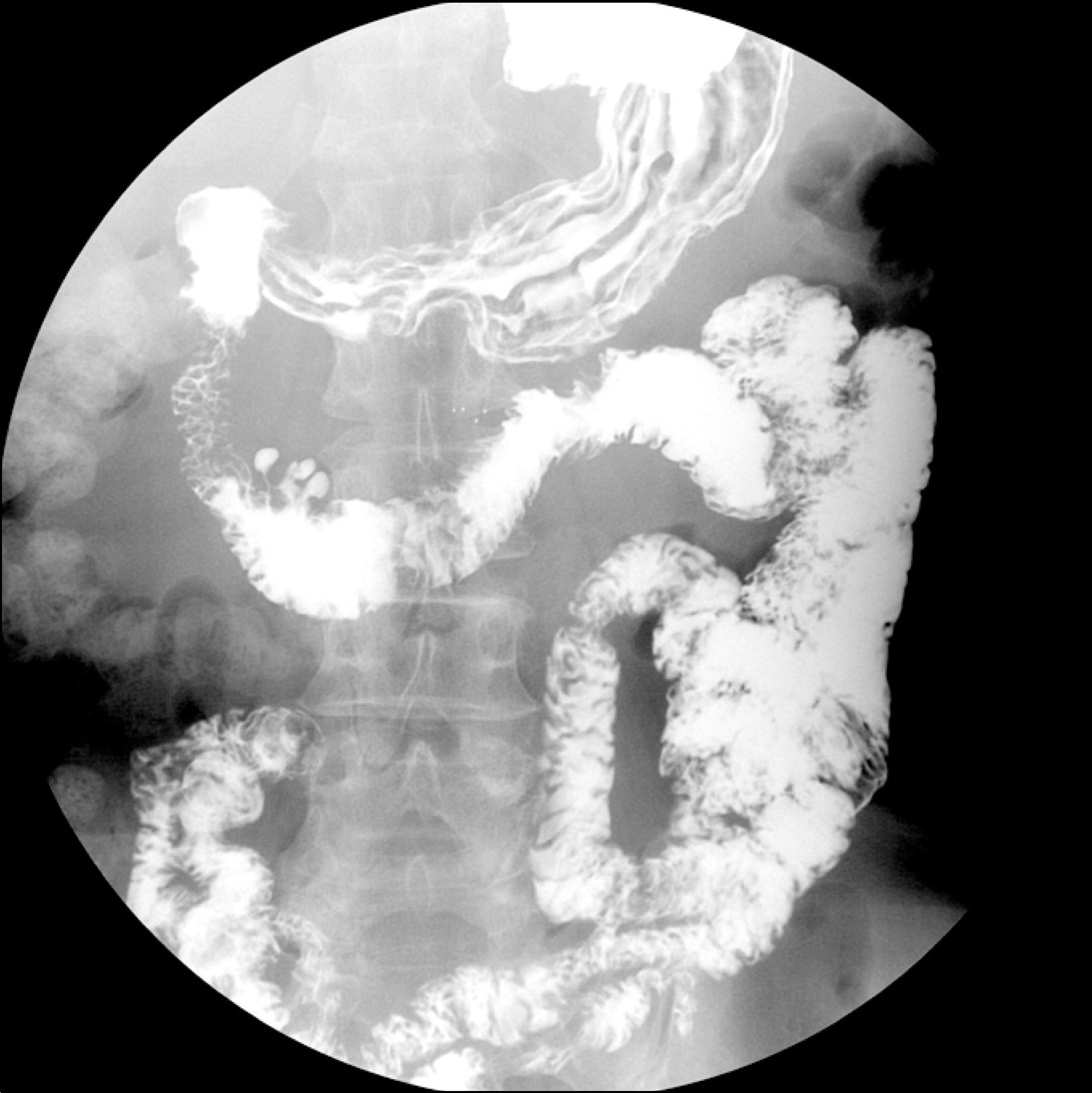
Fig. 3.
Upper gastrointestinal series show no definite evidence of luminal narrowing or passage disturbance.