Abstract
Background/Aims
Few studies have assessed the prognostic value of the primary tumor maximum standardized uptake value (SUVmax) measured by 2-[18 F]-fluoro-2-deoxy-D-glucose PET-CT for patients with bile duct and gallbladder cancer.
Methods
A retrospective analysis of 61 patients with confirmed bile duct and gallbladder cancer who underwent FDG PET-CT in Kangbuk Samsung Medical Center (Seoul, Korea) from April 2008 to April 2011. Prognostic significance of SUVmax and other clinicopathological variables was assessed.
Results
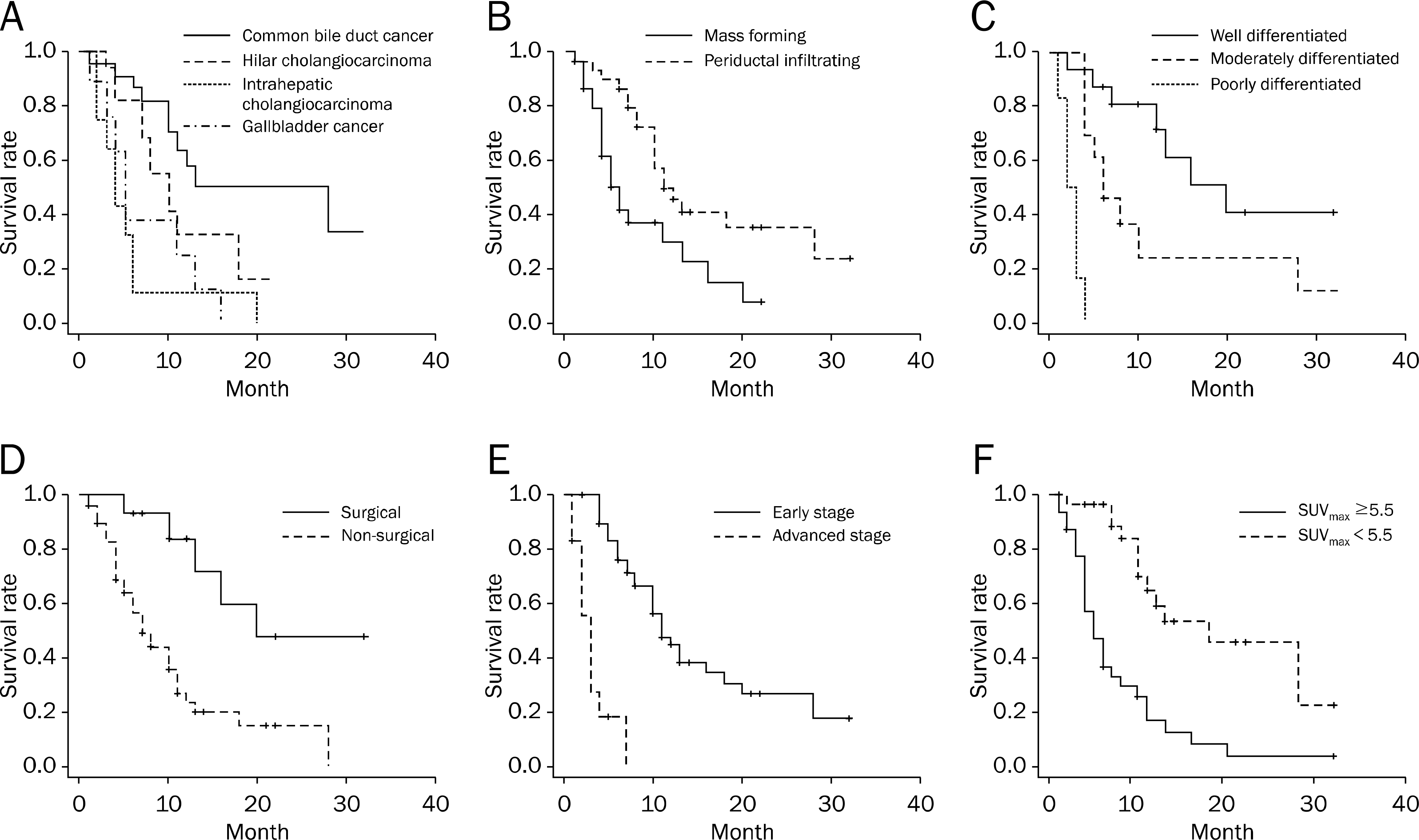
Twenty-three patients were diagnosed as common bile duct cancer, 17 as hilar bile duct cancer, 12 as intrahepatic bile duct cancer, and nine as gallbladder cancer. In univariate analysis, diagnosis of intrahepatic cholangiocarcinoma and gallbladder cancer, mass forming type, poorly differentiated cell type, nonsurgical treatment, advanced American Joint Committee on Cancer (AJCC) staging and primary tumor SUVmax were significant predictors of poor overall survival. In multivariate analysis adjusted for age and sex, primary tumor SUVmax (hazard ratio [HR], 4.526; 95% CI, 1.813–11.299), advanced AJCC staging (HR, 4.843; 95% CI, 1.760–13.328), and nonsurgical treatment (HR, 6.029; 95% CI, 1.989–18.271) were independently associated with poor overall survival.
Go to : 
References
1. Okuda K, Nakanuma Y, Miyazaki M. Cholangiocarcinoma: recent progress. Part 1: epidemiology and etiology. J Gastroenterol Hepatol. 2002; 17:1049–1055.

2. Farley DR, Weaver AL, Nagorney DM. “Natural history” of unresected cholangiocarcinoma: patient outcome after non-curative intervention. Mayo Clinic Proc. 1995; 70:425–429.

3. Park J, Kim MH, Kim KP, et al. Natural history and prognostic factors of advanced cholangiocarcinoma without surgery, chemotherapy, or radiotherapy: a large-scale observational study. Gut Liver. 2009; 3:298–305.

4. Miyazaki M, Ito H, Nakagawa K, et al. Does aggressive surgical resection improve the outcome in advanced gallbladder carcinoma? Hepatogastroenterology. 1999; 46:2128–2132.
5. Jarnagin WR, Fong Y, DeMatteo RP, et al. Staging, resectability, and outcome in 225 patients with hilar cholangiocarcinoma. Ann Surg. 2001; 234:507–517. discussion 517–519.

6. Endo I, Gonen M, Yopp AC, et al. Intrahepatic cholangiocarcinoma: rising frequency, improved survival, and determinants of outcome after resection. Ann Surg. 2008; 248:84–96.
7. Ercolani G, Vetrone G, Grazi GL, et al. Intrahepatic cholangiocarcinoma: primary liver resection and aggressive multimodal treatment of recurrence significantly prolong survival. Ann Surg. 2010; 252:107–114.
8. Petrowsky H, Wildbrett P, Husarik DB, et al. Impact of integrated positron emission tomography and computed tomography on staging and management of gallbladder cancer and cholangiocarcinoma. J Hepatol. 2006; 45:43–50.

9. Kim JY, Kim MH, Lee TY, et al. Clinical role of 18F-FDG PET-CT in suspected and potentially operable cholangiocarcinoma: a prospective study compared with conventional imaging. Am J Gastroenterol. 2008; 103:1145–1151.
10. Lee SW, Kim HJ, Park JH, et al. Clinical usefulness of 18F-FDG PET-CT for patients with gallbladder cancer and cholangiocarcinoma. J Gastroenterol. 2010; 45:560–566.

11. Lee YY, Choi CH, Kim CJ, et al. The prognostic significance of the SUVmax (maximum standardized uptake value for F-18 fluorodeoxyglucose) of the cervical tumor in PET imaging for early cervical cancer: preliminary results. Gynecol Oncol. 2009; 115:65–68.

12. Kitajima K, Kita M, Suzuki K, Senda M, Nakamoto Y, Sugimura K. Prognostic significance of SUVmax (maximum standardized uptake value) measured by [18F]FDG PET/CT in endometrial cancer. Eur J Nucl Med Mol Imaging. 2012; 39:840–845.

13. Ahmadzadehfar H, Rodrigues M, Zakavi R, Knoll P, Mirzaei S. Prognostic significance of the standardized uptake value of pre-therapeutic (18)F-FDG PET in patients with malignant lymphoma. Med Oncol. 2011; 28:1570–1576.

14. Furukawa H, Ikuma H, Asakura K, Uesaka K. Prognostic importance of standardized uptake value on F-18 fluorodeoxy-glucose-positron emission tomography in biliary tract carcinoma. J Surg Oncol. 2009; 100:494–499.

15. Kitamura K, Hatano E, Higashi T, et al. Prognostic value of (18)F-fluorodeoxyglucose positron emission tomography in patients with extrahepatic bile duct cancer. J Hepatobiliary Pancreat Sci. 2011; 18:39–46.
16. Seo S, Hatano E, Higashi T, et al. Fluorine-18 fluorodeoxyglucose positron emission tomography predicts lymph node metastasis, P-glycoprotein expression, and recurrence after resection in mass-forming intrahepatic cholangiocarcinoma. Surgery. 2008; 143:769–777.

17. Lim JH. Cholangiocarcinoma: morphologic classification according to growth pattern and imaging findings. AJR Am J Roentgenol. 2003; 181:819–827.

18. Bosman FT, Carneiro F, Hruban RH, et al. WHO classification of tumours of the digestive system. 4th ed.Geneva: World Health Organization;2010.
19. Edge SB, Byrd DR, Compton CC, Fritz AG, Greene FL, Trotti A. AJCC cancer staging manual. 7th ed.New York: Springer;2010.
20. Kiriyama S, Takada T, Strasberg SM, et al. Tokyo Guidelines Revision Committee. TG13 guidelines for diagnosis and severity grading of acute cholangitis (with videos). J Hepatobiliary Pancreat Sci. 2013; 20:24–34.
21. Vansteenkiste JF, Stroobants SG, Dupont PJ, et al. Prognostic importance of the standardized uptake value on (18)F-fluoro-2-de-oxy-glucose-positron emission tomography scan in non-small-cell lung cancer: An analysis of 125 cases. Leuven Lung Cancer Group. J Clin Oncol. 1999; 17:3201–3206.
22. Sasaki R, Komaki R, Macapinlac H, et al. [18F]fluorodeoxyglucose uptake by positron emission tomography predicts outcome of non-small-cell lung cancer. J Clin Oncol. 2005; 23:1136–1143.

23. Oshida M, Uno K, Suzuki M, et al. Predicting the prognoses of breast carcinoma patients with positron emission tomography using 2-deoxy-2-fluoro[18F]-D-glucose. Cancer. 1998; 82:2227–2234.

24. Lee JD, Yang WI, Park YN, et al. Different glucose uptake and glycolytic mechanisms between hepatocellular carcinoma and intrahepatic mass-forming cholangiocarcinoma with increased (18)F-FDG uptake. J Nucl Med. 2005; 46:1753–1759.
25. Furudoi A, Tanaka S, Haruma K, et al. Clinical significance of human erythrocyte glucose transporter 1 expression at the deep-est invasive site of advanced colorectal carcinoma. Oncology. 2001; 60:162–169.

Go to : 
 | Fig. 1.Univariate analysis of the patient population, tumor characteristics, and overall survival. Kaplan-Meier survival curves with log rank comparisons between each group of final diagnosis (A), morphological type (B), histologic grade (C), treatment method (D), American Joint Committee on Cancer stage (E) and primary tumor standardized uptake value (SUVmax) (F). Log rank p-values were <0.05 for each comparison, respectively. |
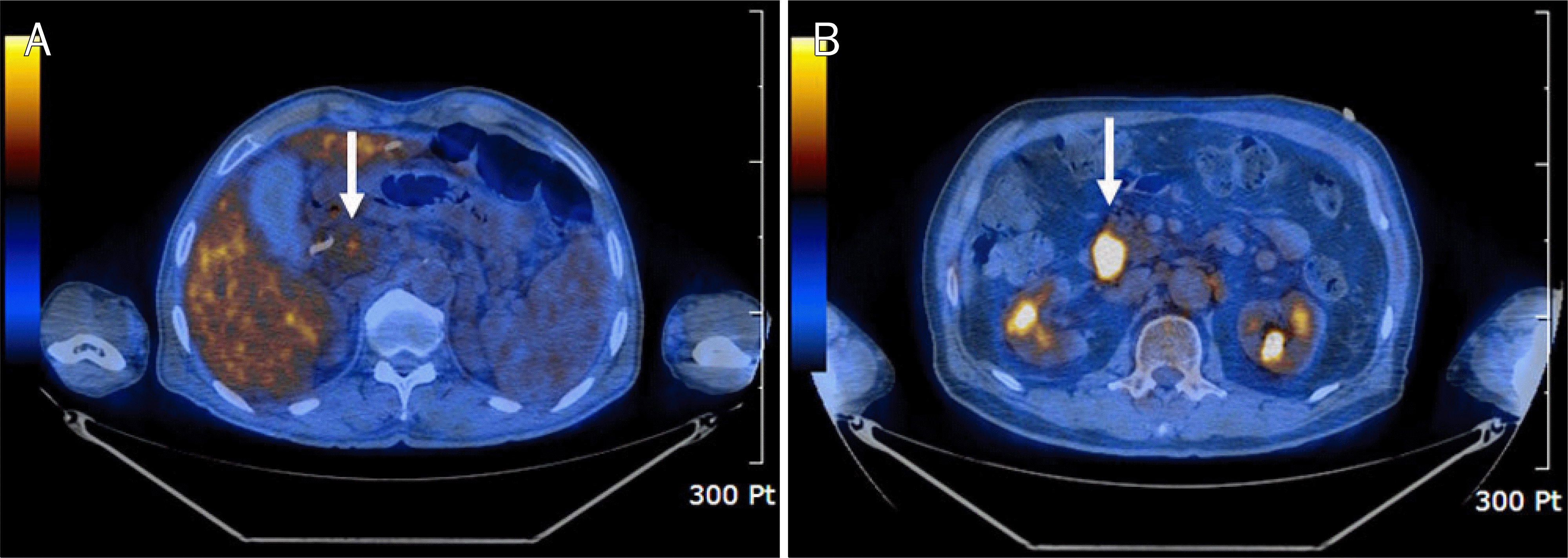
 | Fig. 2.Axial FDG PET-CT images of two patients with common bile duct cancer who received nonsurgical treatment. Primary tumor maximum standardized uptake value is 3.1 (arrow) in 69-year-old man who was alive 26 months after diagnosis (A), and 9.9 (arrow) in 76-year-old man who died 10 months after diagnosis (B), respectively. |
Table 1.
Baseline Clinicopathologic Characteristics of the Patients
Table 2.
Cox Proportional Hazard Regression Analysis of Factors Associated with Overall Survival of Bile Duct and Gallbladder Cancer
| Factor | Patient (n) |
Unadjusted |
Adjusteda |
||||
|---|---|---|---|---|---|---|---|
| HR | 95% CI | p-value | HR | 95% CI | p-value | ||
| Primary tumor SUVmax | |||||||
| <5.5 | 32 | 1 | 1 | ||||
| ≥5.5 | 29 | 3.927 | 1.968–7.836 | <0.001 | 4.526 | 1.813–11.299 | 0.001 |
| AJCC stage | |||||||
| Early (I/II) | 49 | 1 | 1 | ||||
| Advanced (III/IV) | 12 | 10.646 | 4.428–25.596 | <0.001 | 4.843 | 1.760–13.328 | 0.002 |
| Treatment | |||||||
| Surgical | 14 | 1 | 1 | ||||
| Nonsurgical | 47 | 4.053 | 1.559–10.537 | 0.004 | 6.029 | 1.989–18.271 | 0.001 |
| Morphology | |||||||
| Mass forming | 31 | 1 | 1 | ||||
| Periductal infiltrating | 30 | 0.418 | 0.217–0.803 | 0.009 | 0.828 | 0.335–2.050 | 0.684 |
| Age (yr) | |||||||
| ≥65 | 41 | 1 | 1 | ||||
| <65 | 20 | 0.544 | 0.262–1.128 | 0.102 | 1.017 | 0.976–1.061 | 0.417 |
| Sex | |||||||
| Male | 32 | 1 | 1 | ||||
| Female | 29 | 1.424 | 0.744–2.726 | 0.286 | 1.19 | 0.566–2.504 | 0.646 |
| Diagnosis | |||||||
| CBDC | 23 | 1 | |||||
| Hilar | 17 | 2.159 | 0.889–5.242 | 0.089 | |||
| IBDC | 12 | 6.354 | 2.520–16.019 | <0.001 | |||
| GBC | 9 | 4.153 | 1.585–10.883 | 0.004 | |||
| Histology | |||||||
| Well | 16 | 1 | |||||
| Moderate | 13 | 2.651 | 0.979–7.182 | 0.055 | |||
| Poor | 6 | 27.532 | 6.494–116.730 | <0.001 | |||


 PDF
PDF ePub
ePub Citation
 Print
Print


 XML Download