

 PDF
PDF ePub
ePub Citation
 Print
Print


Abstract
Background/Aims
Gastroesophageal reflux disease (GERD) is a common upper gastrointestinal disorder in patients with chronic kidney disease (CKD). However, little is known about the prevalence of GERD in dialysis patients. The aim of the present study was to investigate the difference in the prevalence of GERD in peritoneal dialysis and hemodialysis patients.
Methods
From July 2010 to August 2011, peritoneal dialysis patients (n=30) and hemodialysis patients (n=38) were enrolled. The prevalences of GERD were assessed at a single center with endoscopic findings and interviews using a questionnaire. Also, risk factors of GERD were evaluated.
Results
The prevalences of GERD in peritoneal dialysis and hemodialysis patients were 33.3% and 39.5% (p=0.748), respectively. The prevalences of erosive reflux esophagitis (ERD) in peritoneal dialysis and hemodialysis patients were 16.7% and 23.7% (p=0.477), respectively. The prevalences of nonerosive reflux disease (NERD) in peritoneal dialysis and hemodialysis patients were 16.7% and 13.2% (p=0.685), respectively. The prevalences of GERD, ERD and NERD were higher than those of the general population. The risk factor for GERD was age in hemodialysis patients.
Go to : 
References
1. Jamidar P, Kendall B. Gastrointestinal disease in patients with chronic renal failure. Nissenson AR, Fine RN, Gentile DE, editors. Clinical dialysis. 3rd ed.Norwalk: Appleton and Lange;1995. p. 609–611.
2. Bargman JM. Noninfectious complications of peritoneal dialysis. Gokal R, Nolph KD, editors. The textbook of peritoneal dialysis. 2nd ed.Dordrecht: Kluwer Academic;1994. p. 565.

3. Kim MJ, Kwon KH, Lee SW. Gastroesophageal reflux disease in CAPD patients. Adv Perit Dial. 1998; 14:98–101.
4. Kawaguchi Y, Mine T, Kawana I, et al. Gastroesophageal reflux disease in chronic renal failure patients: evaluation by endoscopic examination. Tokai J Exp Clin Med. 2009; 34:80–83.
5. Yoo SS, Lee WH, Ha J, et al. The prevalence of esophageal disorders in the subjects examined for health screening. Korean J Gastroenterol. 2007; 50:306–312.
6. Oh JH, Choi MG, Kim HR, et al. Clinical spectrum of endoscopic reflux esophagitis in routine check-up subjects in Korea. Korean J Neurogastrointest Motil. 2006; 12:12–18.
7. Na IK, Jung JI, Pard HS. The prevalence and associated factors of reflux esophagitis in routine check-up subjects. J Korean Acad Fam Med. 2001; 22:1647–1655.
8. Kim N, Lee SW, Cho SI, et al. H pylori and Gerd Study Group of Korean College of Helicobacter and Upper Gastrointestinal Research. The prevalence of and risk factors for erosive oesophagitis and nonerosive reflux disease: a nationwide multicentre prospective study in Korea. Aliment Pharmacol Ther. 2008; 27:173–185.
9. Cho YK, Kim GH, Kim JH, Jung HY, Lee JS, Kim NY. Korean Society of Neurogastroenterology and Motility. Diagnosis of gastroesophageal reflux disease: a systematic review. Korean J Gastroenterol. 2010; 55:279–295.
10. Hammer J, Oesterreicher C, Hammer K, Koch U, Traindl O, Kovarik J. Chronic gastrointestinal symptoms in hemodialysis patients. Wien Klin Wochenschr. 1998; 110:287–291.
11. Anderson JE, Yim KB, Crowell MD. Prevalence of gastroesophageal reflux disease in peritoneal dialysis and hemodialysis patients. Adv Perit Dial. 1999; 15:75–78.
12. Twardowski ZJ, Khanna R, Nolph KD, et al. Intraabdominal pressures during natural activities in patients treated with continuous ambulatory peritoneal dialysis. Nephron. 1986; 44:129–135.
13. Hylander BI, Dalton CB, Castell DO, Burkart J, Rossner S. Effect of intraperitoneal fluid volume changes on esophageal pressures: studies in patients on continuous ambulatory peritoneal dialysis. Am J Kidney Dis. 1991; 17:307–310.
14. Lundell LR, Dent J, Bennett JR, et al. Endoscopic assessment of oesophagitis: clinical and functional correlates and further validation of the Los Angeles classification. Gut. 1999; 45:172–180.
15. Kim DM, Ahn CW, Nam SY. Prevalence of obesity in Korea. Obes Rev. 2005; 6:117–121.
16. Van Vlem B, Schoonjans R, Vanholder R, Vandamme W, De Vos M, Lameire N. Dyspepsia and gastric emptying in chronic renal failure patients. Clin Nephrol. 2001; 56:302–307.
17. Rosaida MS, Goh KL. Gastro-oesophageal reflux disease, reflux oesophagitis and nonerosive reflux disease in a multiracial Asian population: a prospective, endoscopy based study. Eur J Gastroenterol Hepatol. 2004; 16:495–501.
18. Yi SY, Lee SK, Kim MH, Han DS, Kim JW, Min YI. Epidemiologic study of reflux esophagitis in general health screening people. Korean J Med. 1994; 46:514–520.
19. Lee SJ, Song CW, Jeen YT, et al. Prevalence of endoscopic reflux esophagitis among Koreans. J Gastroenterol Hepatol. 2001; 16:373–376.
20. Jung SA, Jung HY, Kim KR, Min YI. The prevalence of reflux esophagitis of Korean adults for 10 years of 1990's. Korean J Gastrointest Motil. 2001; 7:161–167.
21. Hwang JK, Kim JH, Hong SG, et al. A prospective multicenter study on the prevalence and symptoms of erosive reflux esophagitis in secondary and tertiary hospitals in Korea. Korean J Gastroenterol. 2009; 53:283–291.
22. Kim BC, Yoon YH, Jyung HS, et al. Clinical characteristics of gastroesophageal reflux diseases and association with Helicobacter pylori infection. Korean J Gastroenterol. 2006; 47:363–369.
23. de Jesús Ventura M, Amato D, Correa-Rotter R, Paniagua R. Relationship between fill volume, intraperitoneal pressure, body size, and subjective discomfort perception in CAPD patients. Mexican Nephrology Collaborative Study Group. Perit Dial Int. 2000; 20:188–193.
24. Pilotto A, Franceschi M, Leandro G, et al. Clinical features of reflux esophagitis in older people: a study of 840 consecutive patients. J Am Geriatr Soc. 2006; 54:1537–1542.
25. Cho YS, Choi MG, Jeong JJ, et al. Prevalence and clinical spectrum of gastroesophageal reflux: a population-based study in Asan-si, Korea. Am J Gastroenterol. 2005; 100:747–753.
26. Fujiwara Y, Higuchi K, Watanabe Y, et al. Prevalence of gastroesophageal reflux disease and gastroesophageal reflux disease symptoms in Japan. J Gastroenterol Hepatol. 2005; 20:26–29.
27. Kwon JH, Chung IS, Son HS, et al. The relationship of gastrin, pepsinogen, and Helicobacter pylori in erosive reflux esophagitis. Korean J Gastroenterol. 2008; 51:159–166.
28. Sanchez NC, Tenofsky PL, Dort JM, Shen LY, Helmer SD, Smith RS. What is normal intraabdominal pressure? Am Surg. 2001; 67:243–248.
29. el-Serag HB, Sonnenberg A. Opposing time trends of peptic ulcer and reflux disease. Gut. 1998; 43:327–333.
30. Asaka M, Sugiyama T, Nobuta A, Kato M, Takeda H, Graham DY. Atrophic gastritis and intestinal metaplasia in Japan: results of a large multicenter study. Helicobacter. 2001; 6:294–299.
31. Gao BX, Duan LP, Wang K, Xia ZW, Lin SR. The roles of Helicobacter pylori and pattern of gastritis in the pathogenesis of reflux esophagitis. Zhonghua Yi Xue Za Zhi. 2006; 86:2674–2678.
32. Masjedizadeh R, Hajiani E, Moezardalan K, et al. H. pylori infection and reflux oesophagitis: a casecontrol study. World J Gastroenterol. 2006; 12:5658–5662.
33. Kim N, Lee SW, Kim JI, et al. H pylori and GERD Study Group of Korean College of Helicobacter and Upper Gastrointestinal Research. Effect of Helicobacter pylori eradication on the development of reflux esophagitis and gastroesophageal reflux symptoms: a nationwide multicenter prospective study. Gut Liver. 2011; 5:437–446.
Go to :
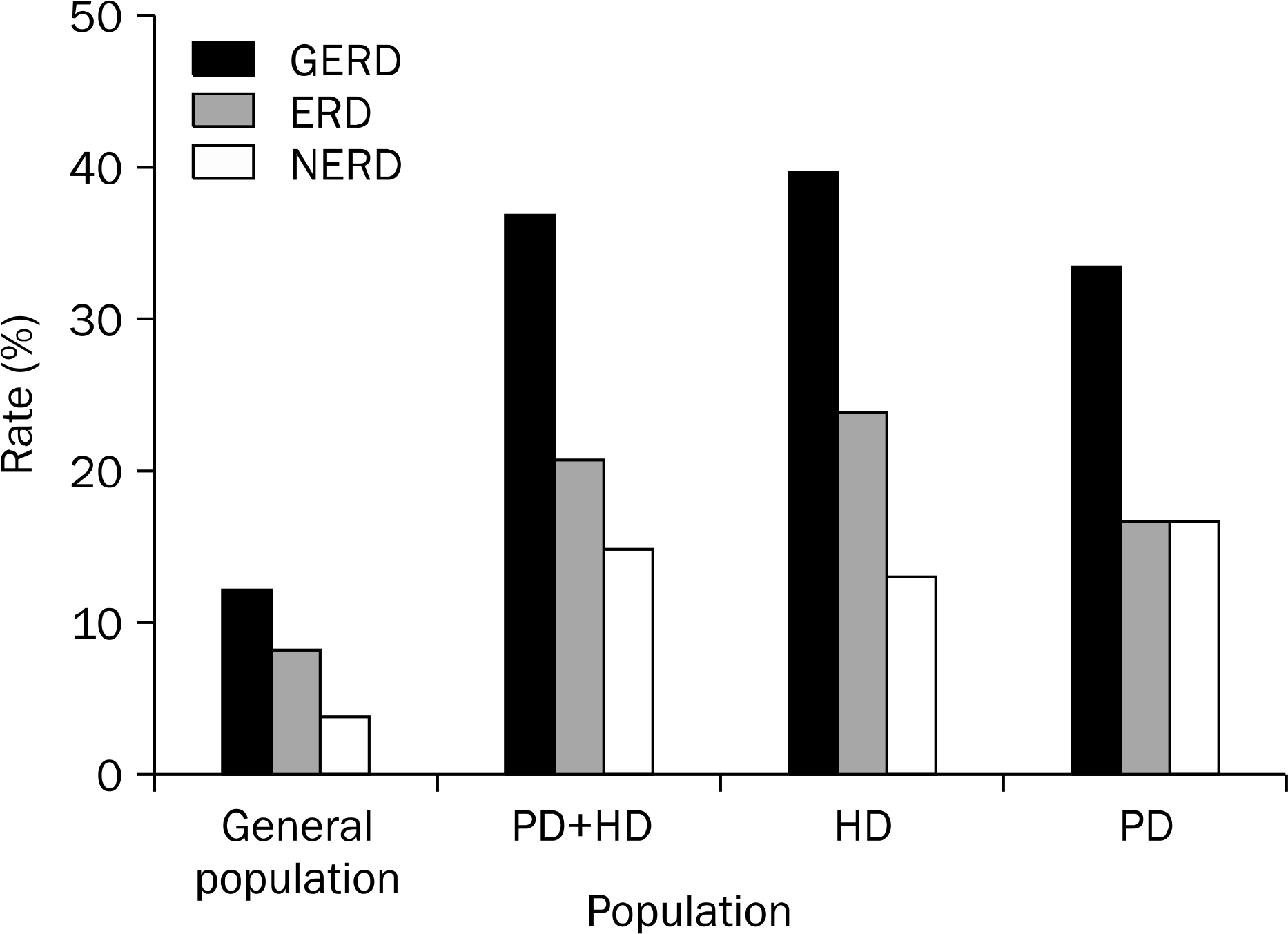 | Fig. 1.Prevalences of gastroesophageal reflux disease (GERD), erosive reflux esophagitis (ERD) and nonerosive reflux disease (NERD). PD, peritoneal dialysis; HD, hemodialysis. The general population data from a nationwide multicenter prospective study in Korea abstracted from the article of Kim et al.8
|
Table 1.
Clinical Characteristics
Table 2.
Factors Associated with Gastroesophageal Reflux Disease in Dialysis Patients by Univariate Analysis
Table 3.
Factors Associated with Gastroesophageal Reflux Disease in Peritoneal Dialysis Patients by Univariate Analysis
Table 4.
Factors Associated with Gastroesophageal Reflux Disease in Hemodialysis Patients by Univariate Analysis
 XML Download