

 PDF
PDF ePub
ePub Citation
 Print
Print


Abstract
Because of its safety and treatment effectiveness, the popularity of radiofrequency ablation (RFA) for the treatment of hepatocellular carcinoma (HCC) has gradually increased. However, some serious complications of RFA such as hepatic infarction, bowel perforation, and tumor seeding have been reported. Recently, we experienced a case of diaphragmatic hernia after RFA for HCC. A 61-year-old man with alcoholic cirrhosis was diagnosed with a 1.0 cm sized HCC in segment (S) 5 and a 1.3 cm sized HCC in S 8 of the liver. He was treated by transarterial chemoembolization and RFA. After RFA, an abdominal CT revealed a diaphragmatic defect with herniating mesentery. Twenty-two months after the RFA, the chest CT showed the diaphragmatic defect with herniating colon and mesentery. Because he had no symptoms, and surgical repair for the diaphragmatic hernia would be a high risk operation for him, we decided to treat the patient conservatively. For its great rarity, we report this case with a review of the literature.
References
1. Rossi S, Di Stasi M, Buscarini E, et al. Percutaneous RF interstitial thermal ablation in the treatment of hepatic cancer. AJR Am J Roentgenol. 1996; 167:759–768.

2. Yan K, Chen MH, Yang W, et al. Radiofrequency ablation of hepatocellular carcinoma: long-term outcome and prognostic factors. Eur J Radiol. 2008; 67:336–347.
3. Bertot LC, Sato M, Tateishi R, Yoshida H, Koike K. Mortality and complication rates of percutaneous ablative techniques for the treatment of liver tumors: a systematic review. Eur Radiol. 2011; 21:2584–2596.
4. Park SG, Park SJ, Koo HS, et al. Biliary-duodenal fistula following radiofrequency ablation therapy for hepatocellular carcinoma. Korean J Gastroenterol. 2008; 51:199–203.
5. Kim JY, Kwon YH, Lee SJ, et al. Abscesso-colonic fistula following radiofrequency ablation therapy for hepatocellular carcinoma;a case successfully treated with histoacryl embolization. Korean J Gastroenterol. 2011; 58:270–274.
6. Liu JG, Wang YJ, Du Z. Radiofrequency ablation in the treatment of small hepatocellular carcinoma: a meta analysis. World J Gastroenterol. 2010; 16:3450–3456.
7. Guglielmi A, Ruzzenente A, Valdegamberi A, et al. Radiofrequency ablation versus surgical resection for the treatment of hepatocellular carcinoma in cirrhosis. J Gastrointest Surg. 2008; 12:192–198.
8. Goldberg SN, Charboneau JW, Dodd GD 3rd, et al. International Working Group on Image-Guided Tumor Ablation. Image-guided tumor ablation: proposal for standardization of terms and reporting criteria. Radiology. 2003; 228:335–345.
9. Koda M, Ueki M, Maeda N, Murawaki Y. Diaphragmatic perforation and hernia after hepatic radiofrequency ablation. AJR Am J Roentgenol. 2003; 180:1561–1562.
10. Shibuya A, Nakazawa T, Saigenji K, Furuta K, Matsunaga K. Diaphragmatic hernia after radiofrequency ablation therapy for hepatocellular carcinoma. AJR Am J Roentgenol. 2006; 186(5 Suppl):S241–S243.
11. di Francesco F, di Sandro S, Doria C, et al. Diaphragmatic hernia occurring 15 months after percutaneous radiofrequency ablation of a hepatocellular cancer. Am Surg. 2008; 74:129–132.
12. Yamagami T, Yoshimatsu R, Matsushima S, Tanaka O, Miura H, Nishimura T. Diaphragmatic hernia after radiofrequency ablation for hepatocellular carcinoma. Cardiovasc Intervent Radiol. 2011; 34(Suppl 2):S175–S177.
13. Singh M, Singh G, Pandey A, Cha CH, Kulkarni S. Laparoscopic repair of iatrogenic diaphragmatic hernia following radiofrequency ablation for hepatocellular carcinoma. Hepatol Res. 2011; 41:1132–1136.
14. Tateishi R, Shiina S, Teratani T, et al. Percutaneous radiofrequency ablation for hepatocellular carcinoma. An analysis of 1000 cases. Cancer. 2005; 103:1201–1209.
15. Curley SA, Izzo F, Ellis LM, Nicolas Vauthey J, Vallone P. Radiofrequency ablation of hepatocellular cancer in 110 patients with cirrhosis. Ann Surg. 2000; 232:381–391.
16. Uehara T, Hirooka M, Ishida K, et al. Percutaneous ultrasound-guided radiofrequency ablation of hepatocellular carcinoma with artificially induced pleural effusion and ascites. J Gastroenterol. 2007; 42:306–311.
17. Raman SS, Aziz D, Chang X, Sayre J, Lassman C, Lu D. Minimizing diaphragmatic injury during radiofrequency ablation: efficacy of intraabdominal carbon dioxide insufflation. AJR Am J Roentgenol. 2004; 183:197–200.
18. Lee SD, Han HS, Cho JY, et al. Safety and efficacy of laparoscopic radiofrequency ablation for hepatic malignancies. J Korean Surg Soc. 2012; 83:36–42.
19. Iwai S, Sakaguchi H, Fujii H, et al. Benefits of artificially induced pleural effusion and/or ascites for percutaneous radiofrequency ablation of hepatocellular carcinoma located on the liver surface and in the hepatic dome. Hepatogastroenterology. 2012; 59:546–550.
Fig. 1.
Hepatocellular carcinoma in hepatic dome (segment 8). (A) Axial MRI section showing a slightly enhanced nodule in the hepatic dome (arrow) during the arterial phase. (B) Coronal T1 weighted image showing a hypointense nodule in the hepatic dome (arrow).
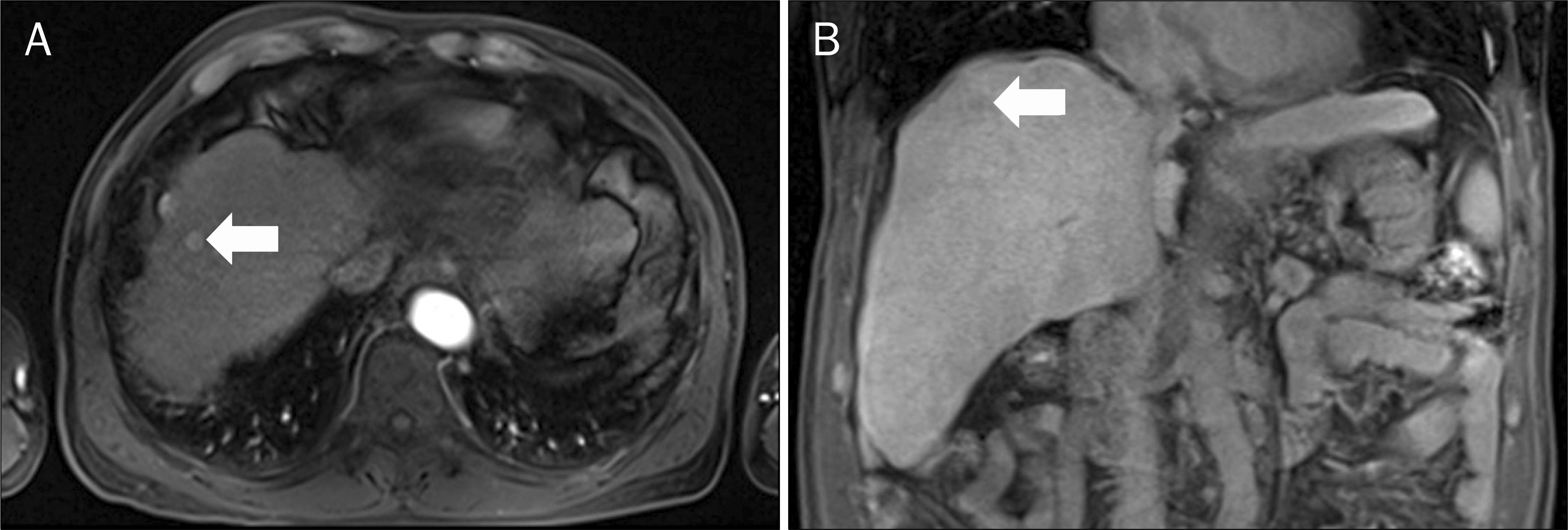
Fig. 2.
Abdomen CT findings. (A) Coronal abdomen CT revealed about a 2.0 cm sized defect (arrowhead) of the diaphragm adjacent to the lesion (arrow) that was ablated. (B) Coronal abdomen CT showed herniation of mesenteric fat and blood vessels (arrow).
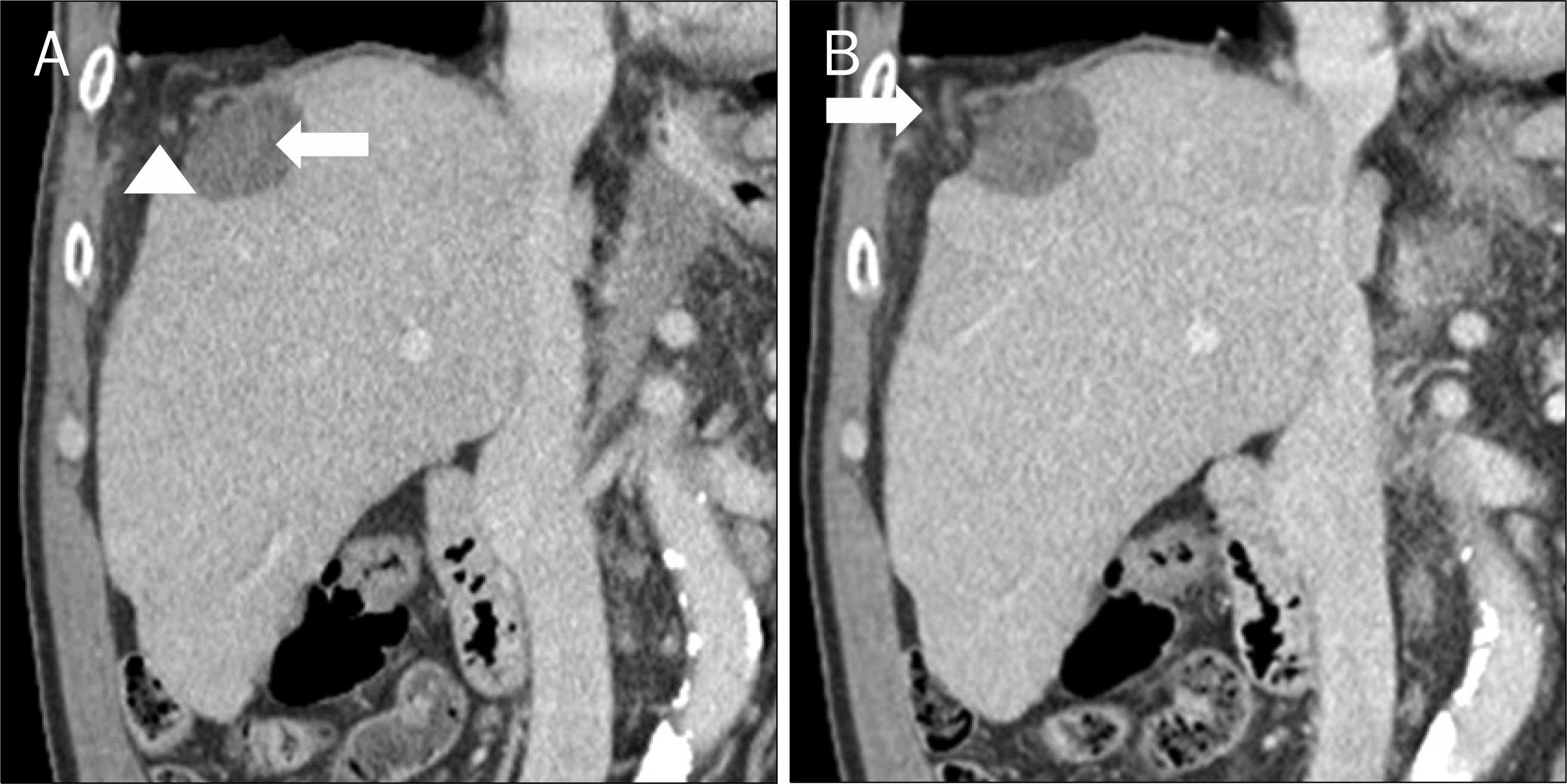
Fig. 3.
Twenty-two months after radiofrequency ablation. (A) Chest X-ray showing a large amount of pleural fluid and loops of bowel (arrow) in the right pleural cavity. (B) Coronal chest CT image showing about a 4.0 cm sized defect of the diaphragm (arrow-head) and herniation of the large intestine (arrow) in the right pleural cavity.
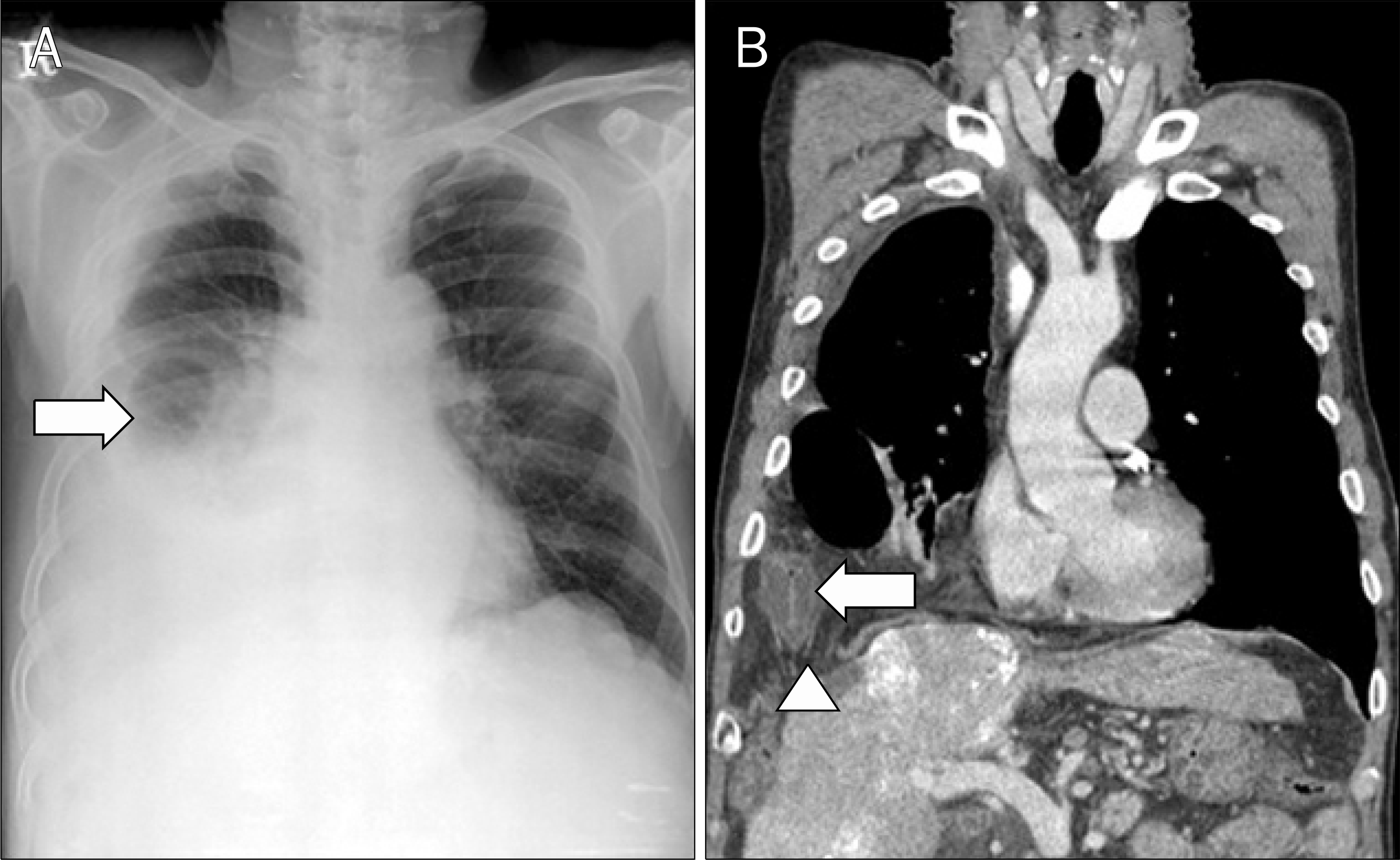
Table 1.
Summary of the Literature of Diaphragmatic Injury Following Radiofrequency Ablation for Hepatocellular Carcinoma
 XML Download