

 PDF
PDF ePub
ePub Citation
 Print
Print


Abstract
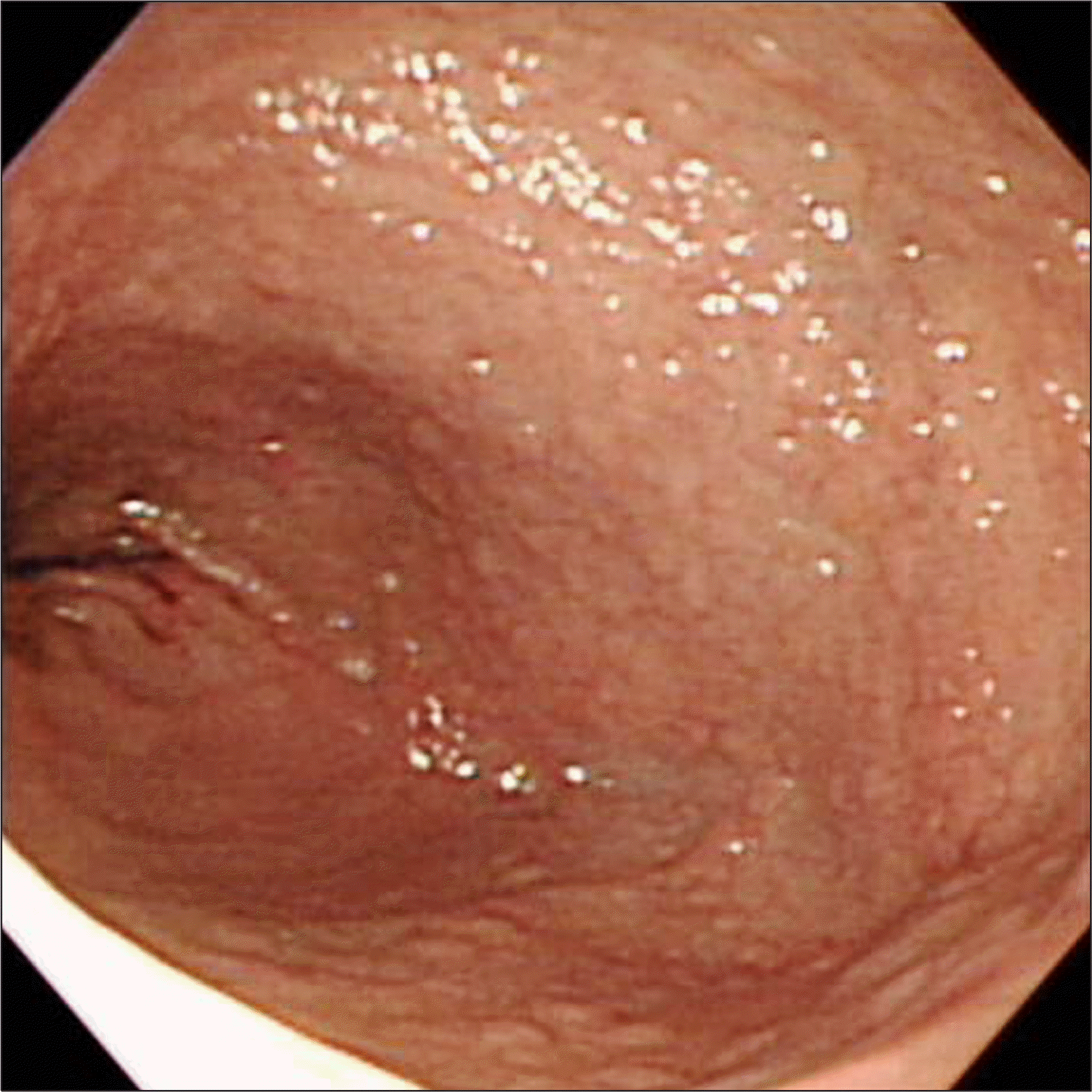
Celiac disease is a chronic absorptive disorder of the small intestine caused by gluten. The prevalence rate of celiac disease is 1% in Western countries. But, it is rare in Asian countries, and there is no celiac disease reported in Korea. Here, we report a case of celiac disease. An 36-years-old woman complained nonspecific abdominal pain and diarrhea. She had anemia and was taking medication for osteoporosis. Colonoscopy showed no abnormality except shallow ulcer at the terminal ileum. Gastroduodenoscopy showed micronodularity at the duodenum 2nd and 3rd portion. Capsule endoscopy and enteroscopy showed villous atrophy and blunting of villi from the duodenum. Small intestinal pathology showed villous atrophy with lymphocyte infiltration. After gluten free diet, diarrhea, abdominal pain, anemia and osteoporosis were improved. And, she felt well-being sensation. This is a first case of celiac disease in Korea.
Go to : 
References
1. Mäki M, Mustalahti K, Kokkonen J, et al. Prevalence of celiac disease among children in Finland. N Engl J Med. 2003; 348:2517–2524.

2. Rubio-Tapia A, Ludvigsson JF, Brantner TL, Murray JA, Everhart JE. The prevalence of celiac disease in the United States. Am J Gastroenterol. 2012; 107:1538–1544.
3. Corazza GR, Di Sario A, Cecchetti L, et al. Bone mass and metabolism in patients with celiac disease. Gastroenterology. 1995; 109:122–128.
4. Kemppainen T, Kröger H, Janatuinen E, et al. Osteoporosis in adult patients with celiac disease. Bone. 1999; 24:249–255.
5. James SP. National Institutes of Health Consensus Development Conference Statement on Celiac Disease, June 28–30, 2004. Gastroenterology. 2005; 128(4 Suppl 1):S1–S9.
6. Rostom A, Murray JA, Kagnoff MF. American Gastroenterological Association (AGA) Institute technical review on the diagnosis and management of celiac disease. Gastroenterology. 2006; 131:1981–2002.
7. Armstrong MJ, Hegade VS, Robins G. Advances in coeliac disease. Curr Opin Gastroenterol. 2012; 28:104–112.
8. Cummins AG, Roberts-Thomson IC. Prevalence of celiac disease in the Asia-Pacific region. J Gastroenterol Hepatol. 2009; 24:1347–1351.
Go to :
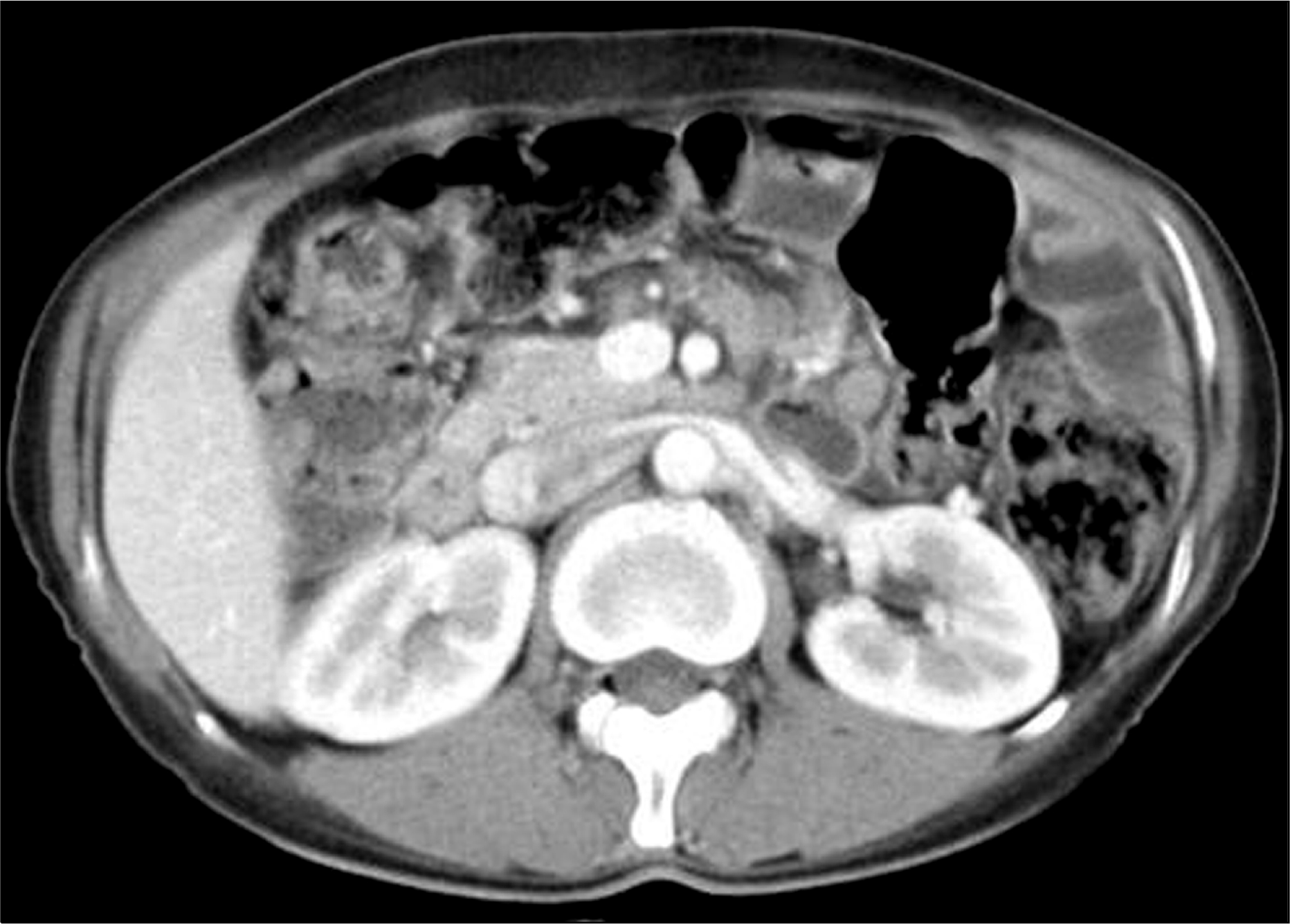 | Fig. 2.Abdominal CT finding. Abdominal CT showed multiple conglomerated lymphadenopathies in the mesentery. |
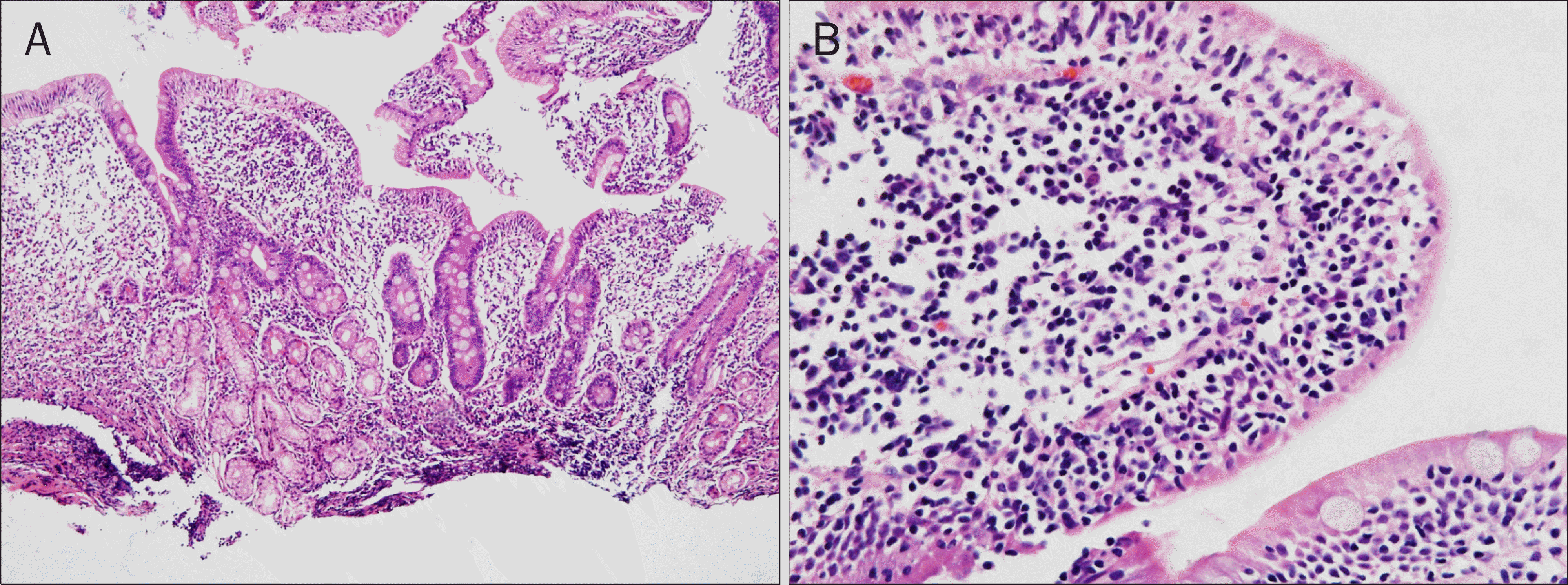 | Fig. 4.Microscopic findings of the duodenal 2nd portion. (A) Villous atrophy, crypt hyperplasia in a biopsy of duodenum 2nd portion (H&E, ×100).(B) Increase in the intraepithelial lymphocytes was noted (H&E, ×400). |
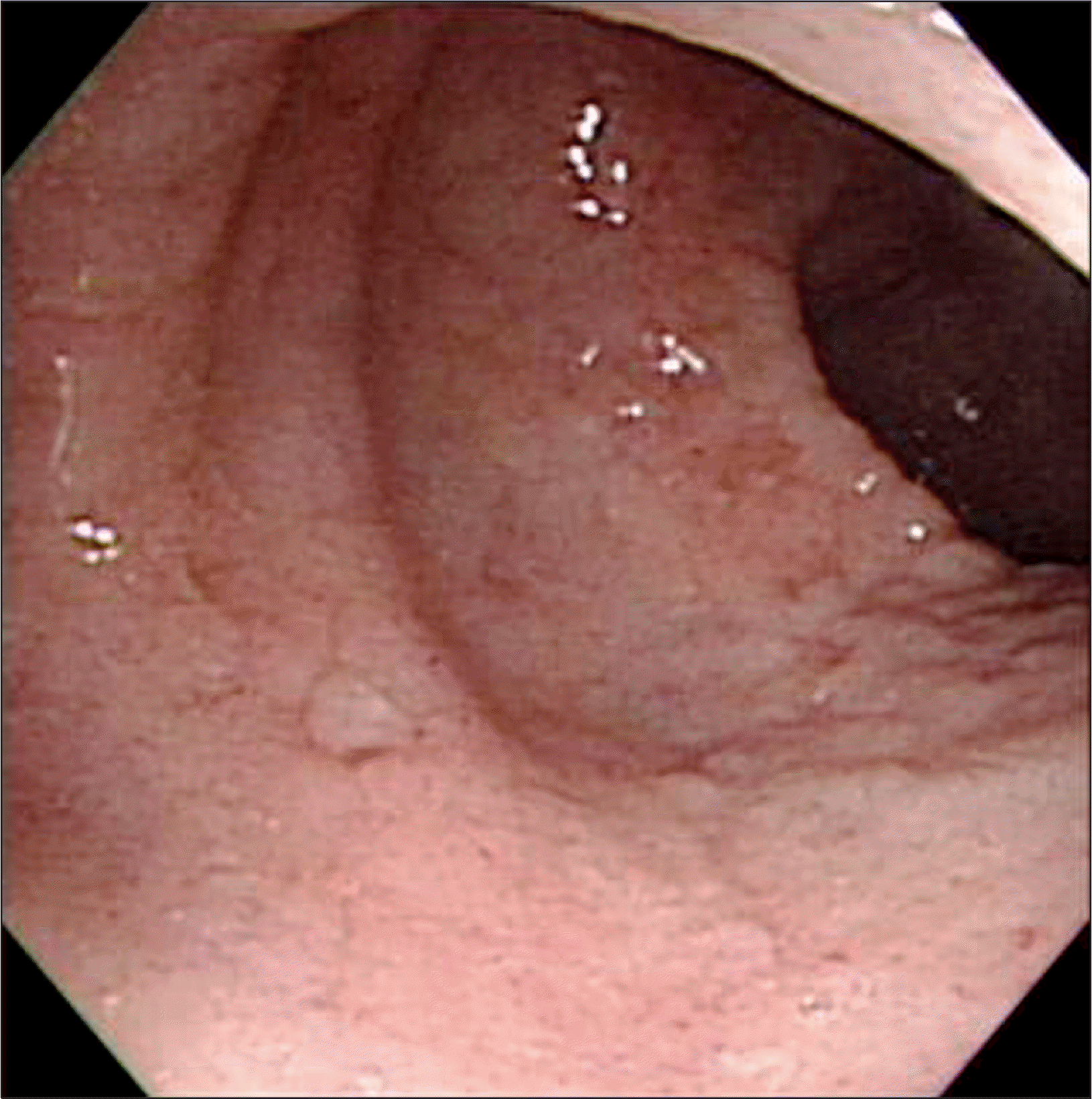
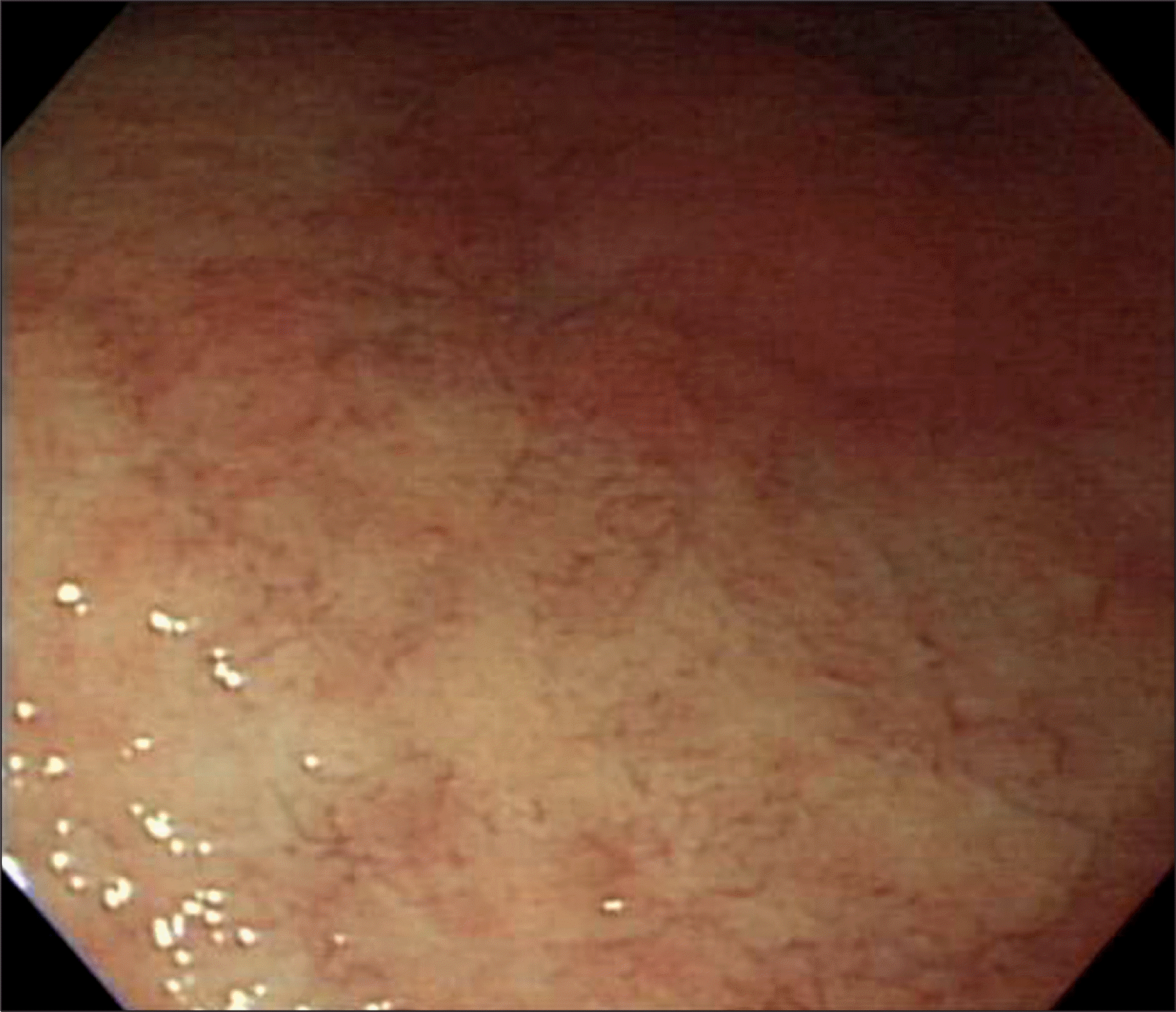
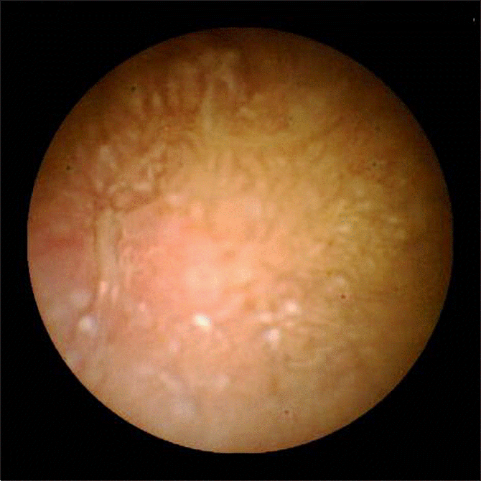 | Fig. 5.Capsule endoscopic finding. It showed blunted villi with focal atrophic change at the jejunum. |

 XML Download