

 PDF
PDF ePub
ePub Citation
 Print
Print


Abstract
Zollinger-Ellison syndrome (ZES) is characterized by gastrinoma and resultant hypergastrinemia, which leads to recurrent peptic ulcers. Because gastrinoma is the most common pancreatic endocrine tumor seen in multiple endocrine neoplasia type I (MEN 1), the possibility of gastrinoma should be investigated carefully when patients exhibit symptoms associated with hormonal changes. Ureteral stones associated with hyperparathyroidism in the early course of MEN 1 are known to be its most common clinical manifestation; appropriate evaluation and close follow-up of patients with hypercalcemic urolithiasis can lead to an early diagnosis of gastrinoma. We report a patient with ZES associated with MEN 1, and urolithiasis as the presenting entity. A 51-year-old man visited the emergency department with recurrent epigastric pain. He had a history of calcium urinary stone 3 years ago, and 2 years later he had 2 operations for multiple jejunal ulcer perforations; these surgeries were 9 months apart. He was taking intermittent courses of antiulcer medication. Multiple peripancreatic nodular masses, a hepatic metastasis, parathyroid hyperplasia, and a pituitary microadenoma were confirmed by multimodal imaging studies. We diagnosed ZES with MEN 1 and performed sequential surgical excision of the gastrinomas and the parathyroid adenoma. The patient received octreotide injection therapy and close follow-up.
Go to : 
References
1. Vasquez CJ, Gagel RF. General features of gastrointestinal (GI) neuroendocrine tumors. Longo DL, Fauci AS, Kasper DL, Hauser SL, Jameson J, Loscalzo J, editors. Harrison's principles of internal medicine. Volume 1. 18th ed.New York, London: McGraw-Hill Medical;2012. p. 3072–3081.
2. Jensen RT, Norton JA. Endocrine tumors of the pancreas and gastrointestinal tract. Feldman M, Friedman LS, Brandt LJ, editors. Sleisenger and Fordtran's gastrointestinal and liver disease: pathophysiology, diagnosis, management. Volume 1. 9th ed.Philadelphia, London: W.B. Saunders;2010. p. 491–522.

3. Bringhurst FR, Demay MB, Kronenberg HM. Hormones and disorders of mineral metabolism. Melmed S, Polonsky KS, Larsen PR, Kronenberg HM, editors. Williams textbook of endocrinology. 12th ed.Philadelphia: Elsevier;2011. p. 1237–1304.
4. Osefo N, Ito T, Jensen RT. Gastric acid hypersecretory states: recent insights and advances. Curr Gastroenterol Rep. 2009; 11:433–441.
5. Lee YM, Jung HY, Choi SY, et al. A case of metachronous multiple endocrine neoplasia type I manifested as Zollinger-Ellison syndrome. Korean J Gastroenterol. 2002; 39:50–54.
6. Christopoulos C, Antoniou N, Thempeyioti A, Calender A, Economopoulos P. Familial multiple endocrine neoplasia type I: the urologist is first on the scene. BJU Int. 2005; 96:884–887.
7. Yamazaki M, Suzuki S, Kosugi S, et al. MEN Consortium of Japan. Delay in the diagnosis of multiple endocrine neoplasia type 1: typical symptoms are frequently overlooked. Endocr J. 2012; 59:797–807.
8. Wong H, Yau T, Chan P, et al. PPI-delayed diagnosis of gastrinoma: oncologic victim of pharmacologic success. Pathol Oncol Res. 2010; 16:87–91.
9. Corleto VD, Annibale B, Gibril F, et al. Does the widespread use of proton pump inhibitors mask, complicate and/or delay the diagnosis of Zollinger-Ellison syndrome? Aliment Pharmacol Ther. 2001; 15:1555–1561.
10. Tonelli F, Giudici F, Fratini G, Brandi ML. Pancreatic endocrine tumors in multiple endocrine neoplasia type 1 syndrome: review of literature. Endocr Pract. 2011; 17(Suppl 3):33–40.
11. Imamura M. Recent standardization of treatment strategy for pancreatic neuroendocrine tumors. World J Gastroenterol. 2010; 16:4519–4525.
12. Tsalis K, Vrakas G, Vradelis S, et al. Primary hepatic gastrinoma: report of a case and review of literature. World J Gastrointest Pathophysiol. 2011; 2:26–30.
13. Jung DJ, Ha HK, Kim PN, Lee MG, Auh YH. Zollinger-Ellison syndrome: a case report. J Korean Radiol Soc. 1999; 41:1173–1176.
14. Tonelli F, Fratini G, Nesi G, et al. Pancreatectomy in multiple endocrine neoplasia type 1-related gastrinomas and pancreatic endocrine neoplasias. Ann Surg. 2006; 244:61–70.
15. Tomassetti P, Campana D, Piscitelli L, et al. Treatment of Zollinger-Ellison syndrome. World J Gastroenterol. 2005; 11:5423–5432.
Go to :
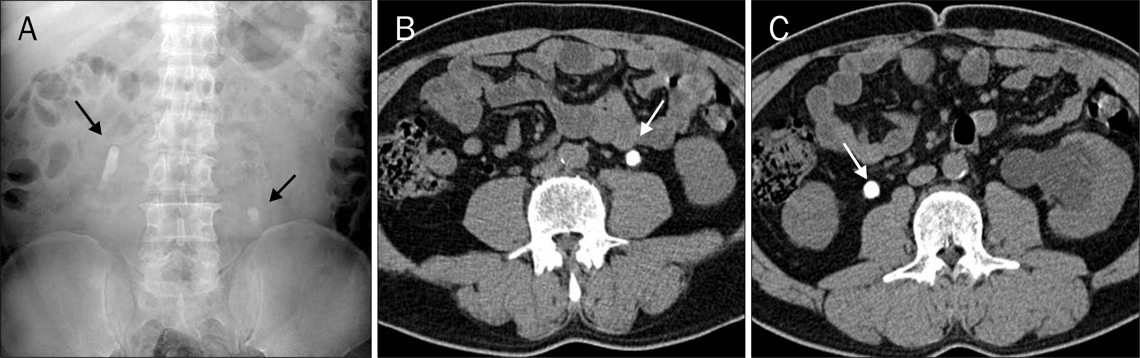 | Fig. 1.Clinical manifestation findings. (A) X-ray examination of the kidneys, ureter, and bladder showed calcified stones (arrows) in both ureters.(B, C) Abdominal nonenhanced CT showed calcified stones (arrows) in both ureters. |
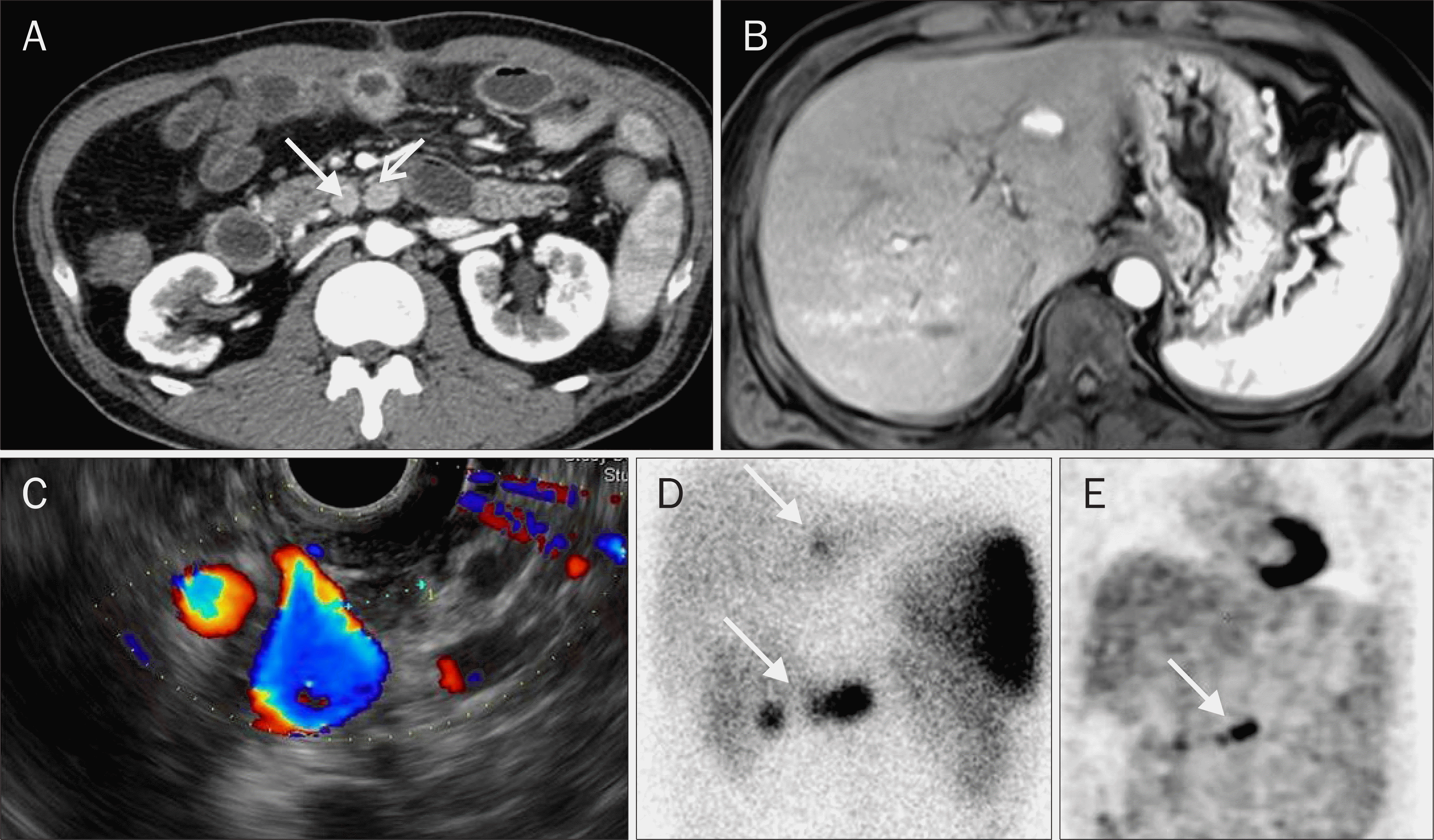 | Fig. 2.Pancreatic gastrinoma findings. (A) Abdominal CT showed multiple restricted nodular masses (arrows). (B) Liver MRI showed hypersignal enhanced lesion in the superior segment of the left lobe. (C) EUS showed well defined hypoechoic nodular lesion. (D) Octerotide scan showed focal hyperactivity in the pancreas head and body portion and mild focal hyperactivity in the left hepatic lobe (arrows). (E) PET-CT showed 18F 2-fluoro-2-deoxyglucose (FDG) hot uptake in pancreatic area (arrow). |
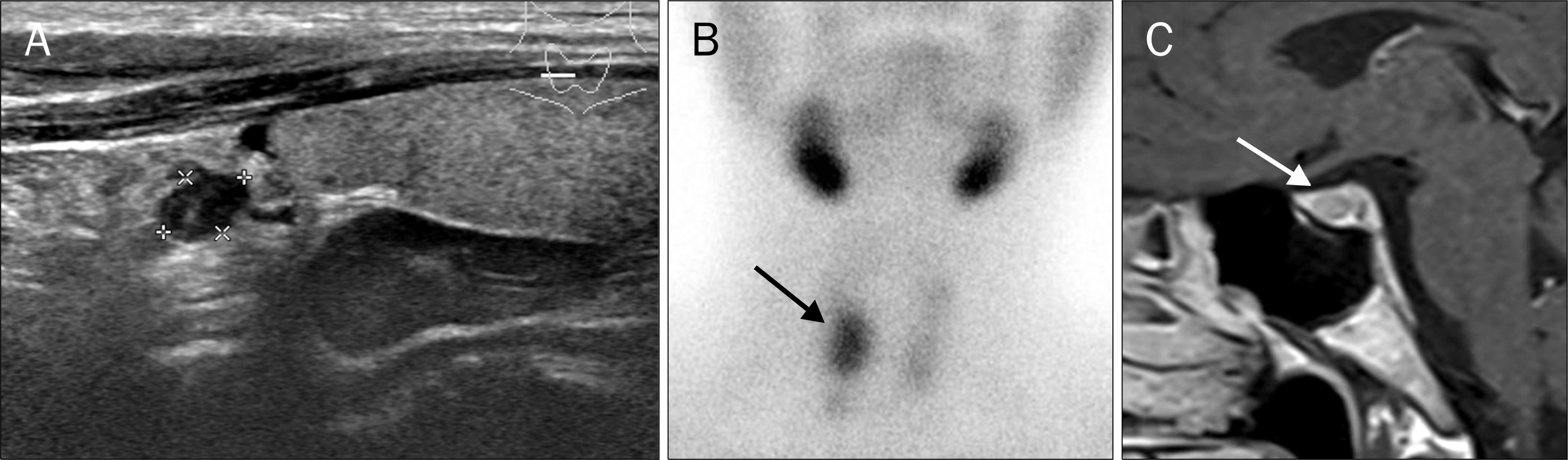 | Fig. 3.Multiple endocrine neoplasia type I findings. (A) Thyroid ultrasonography showed parathyroid adenoma in right superior pole of thyroid.(B) Parathyroid Tc-99m methoxy isobutyl isonitrile scan showed persistent tracer in right thyroid lobe (arrow; after 30 minutes, 60 minutes). (C) Brain MRI showed low attenuation of pituitary such as microadenoma (arrow). |
 XML Download