

 PDF
PDF ePub
ePub Citation
 Print
Print


Abstract
Background/Aims
Autoimmune pancreatitis (AIP) often occurs with obstructive jaundice in old age in cases of weight loss, mimicking pancreatobiliary cancer. This study aimed to determine the sensitivity and specificity serum IgG, IgG4 and CEA, CA 19–9 levels for the diagnosis of AIP and their ability to distinguish AIP from pancreatobiliary cancer.
Methods
The level of serums IgG, IgG4 and CEA, CA 19–9 were measured in 413 patients including 125 with AIP, 201 with pancreatic cancer, and 87 with cholangiocarcinoma.
Results
Among AIP patients, 43.2% (54/125) showed elevated IgG levels (≥1,800 mg/dL) and 52% (65/125) showed elevated IgG4 levels (≥135 mg/dL). Sensitivity and specificity of elevated serum IgG for diagnosis AIP were 43% and 88% respectively, and 52% and 97%, respectively for elevated serum IgG4. When the cut-off value of serum IgG4 was raised to 270 mg/dL (twice the upper limit of normal), the specificity improved to 100%. About 25% of the AIP patients showed an increased level of CA 19–9 at >37 U/mL and about 12.2% of them showed an increased level of CA 19–9 at >100 U/mL. On the contrary, only 1.8% of the AIP patients showed an increased level of CEA at >6.0 ng/mL.
Conclusions
To avoid unnecessary surgeries resulting from a misdiagnosed pancreatobiliary cancer as opposed to AIP, it is necessary to consider both serum immunoglobulin and tumor marker. In particular, because high level of IgG4 (≥270 mg/dL) and CA19–9 (>100 U/mL) are relatively rare in pancreatobiliary cancer and AIP, respectively, they will be helpful in differential diagnosis.
Go to : 
References
1. Kim KP, Kim MH, Song MH, Lee SS, Seo DW, Lee SK. Autoimmune chronic pancreatitis. Am J Gastroenterol. 2004; 99:1605–1616.

2. Yoshida K, Toki F, Takeuchi T, Watanabe S, Shiratori K, Hayashi N. Chronic pancreatitis caused by an autoimmune abnormality. Proposal of the concept of autoimmune pancreatitis. Dig Dis Sci. 1995; 40:1561–1568.
3. Chari ST, Smyrk TC, Levy MJ, et al. Diagnosis of autoimmune pancreatitis: the Mayo Clinic experience. Clin Gastroenterol Hepatol. 2006; 4:1010–1016. quiz 1934.
4. Kim MH, Kwon S. Diagnostic criteria for autoimmune chronic pancreatitis. J Gastroenterol. 2007; 42(Suppl 18):42–49.
5. Okazaki K, Kawa S, Kamisawa T, et al. Research Committee of Intractable Diseases of the Pancreas. Clinical diagnostic criteria of autoimmune pancreatitis: revised proposal. J Gastroenterol. 2006; 41:626–631.
6. Kamisawa T, Egawa N, Nakajima H, Tsuruta K, Okamoto A, Kamata N. Clinical difficulties in the differentiation of autoimmune pancreatitis and pancreatic carcinoma. Am J Gastroenterol. 2003; 98:2694–2699.
7. Weber SM, Cubukcu-Dimopulo O, Palesty JA, et al. Lymphoplasmacytic sclerosing pancreatitis: inflammatory mimic of pancreatic carcinoma. J Gastrointest Surg. 2003; 7:129–137. discussion 137–139.
8. Park DH, Kim MH, Chari ST. Recent advances in autoimmune pancreatitis. Gut. 2009; 58:1680–1689.
9. Hamano H, Kawa S, Horiuchi A, et al. High serum IgG4 concentrations in patients with sclerosing pancreatitis. N Engl J Med. 2001; 344:732–738.
10. Ghazale A, Chari ST, Smyrk TC, et al. Value of serum IgG4 in the diagnosis of autoimmune pancreatitis and in distinguishing it from pancreatic cancer. Am J Gastroenterol. 2007; 102:1646–1653.
11. Sarles H, Sarles JC, Muratore R, Guien C. Chronic inflammatory sclerosis of the pancreas–an autonomous pancreatic disease? Am J Dig Dis. 1961; 6:688–698.
12. Kim JY, Chang HS, Kim MH, et al. A case of autoimmune chronic pancreatitis improved with oral steroid therapy. Korean J Gastroenterol. 2002; 39:304–308.
13. Choi EK, Kim MH, Lee TY, et al. The sensitivity and specificity of serum immunoglobulin G and immunoglobulin G4 levels in the diagnosis of autoimmune chronic pancreatitis: Korean experience. Pancreas. 2007; 35:156–161.
14. Kang P, Lee KT, Sinn DH, et al. Clinical usefulness of serum immunoglobulin G and G4 level in the diagnosis of autoimmune pancreatitis. Korean J Gastroenterol. 2008; 52:304–309.
15. Song TJ, Kim MH, Moon SH, et al. The combined measurement of total serum IgG and IgG4 may increase diagnostic sensitivity for autoimmune pancreatitis without sacrificing specificity, compared with IgG4 alone. Am J Gastroenterol. 2010; 105:1655–1660.
16. van Gulik TM, Reeders JW, Bosma A, et al. Incidence and clinical findings of benign, inflammatory disease in patients resected for presumed pancreatic head cancer. Gastrointest Endosc. 1997; 46:417–423.
17. Steinberg W. The clinical utility of the CA 19–9 tumor-associated antigen. Am J Gastroenterol. 1990; 85:350–355.
18. Kim HK, Lee CH, Ryoo BL, et al. The significaned of serum CA 19–9 level change after biliary drainge procedures in pancreatic and biliary tract cancer. J Korean Cancer Assoc. 1995; 27:929–935.
19. Chae YS, Kim KS, Lee HS, Lee WJ, Kim BR. Clinical analysis of CA19–9 positive rate of hepatobiliary pancreas disease. J Korean Surg Soc. 2002; 63:227–232.
Go to :
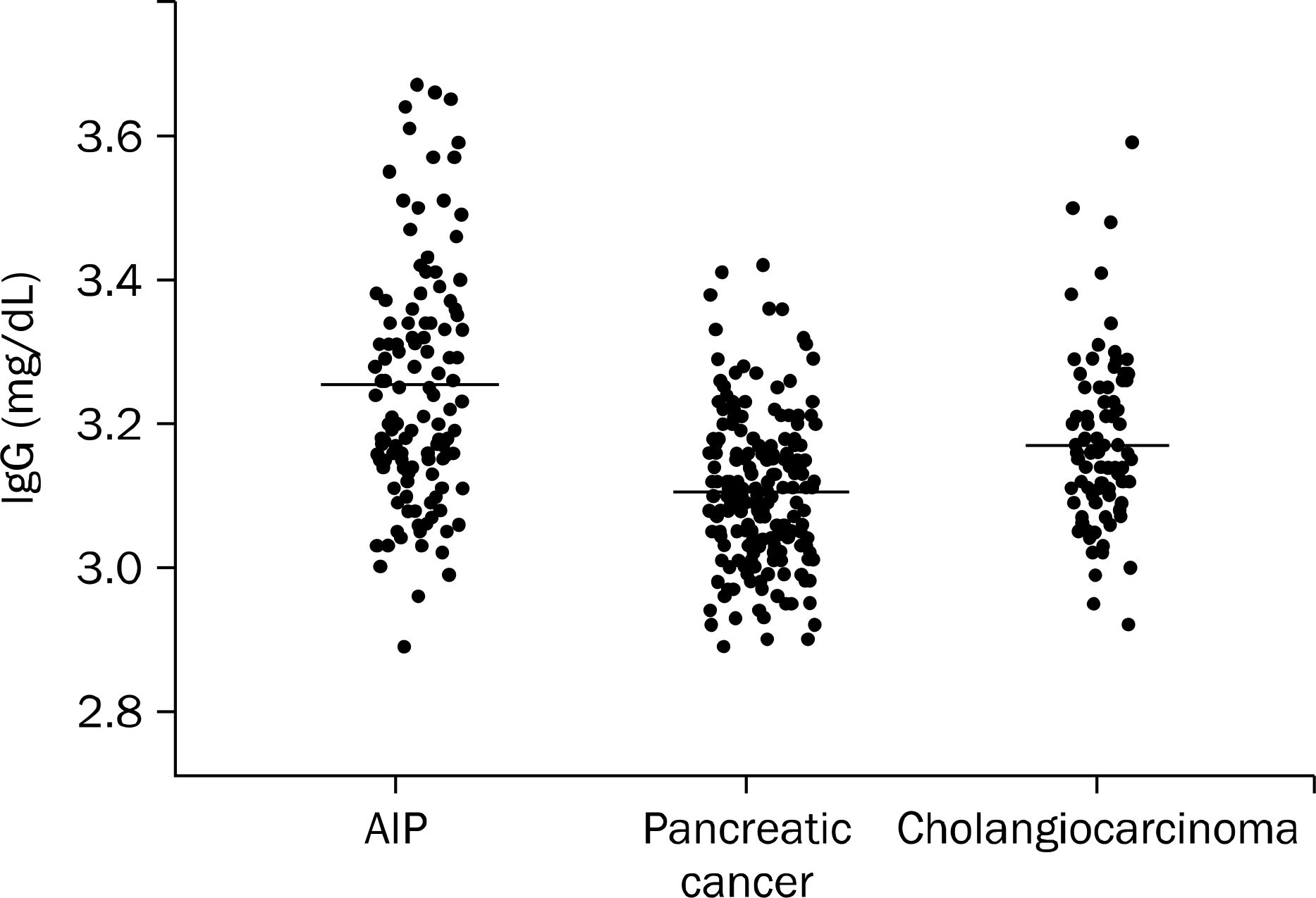 | Fig. 1.Distribution of serum IgG level. There was a significant difference in the serum IgG levels among the 3 groups (Kruskal- Wallis after logarithmic transformation, p<0.001). AIP, autoimmune pancreatitis. |
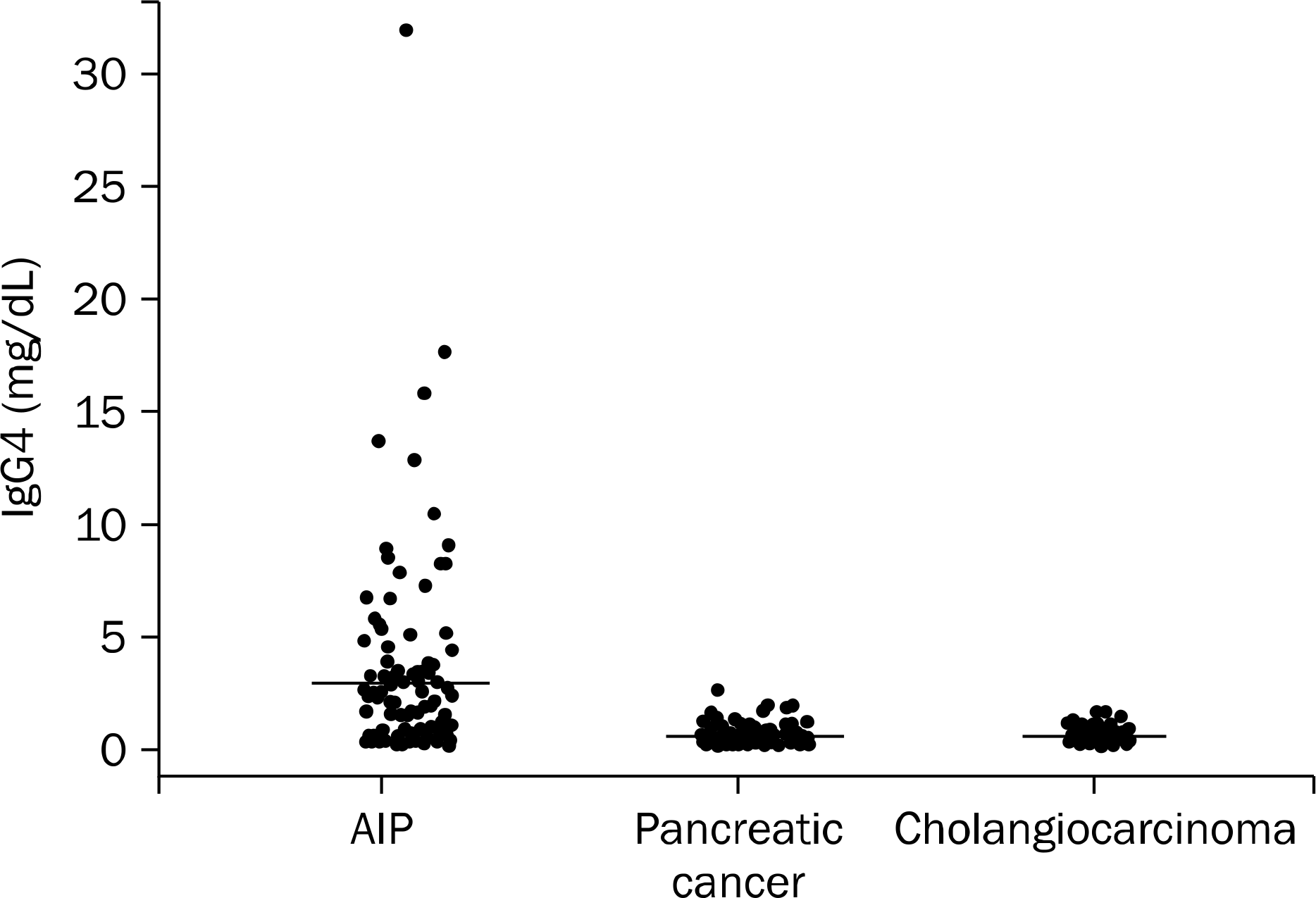 | Fig. 2.Distribution of serum IgG4 level. There was a significant difference in the serum IgG4 levels among the 3 groups (Kruskal- Wallis after logarithmic transformation, p<0.001). AIP, autoimmune pancreatitis. |
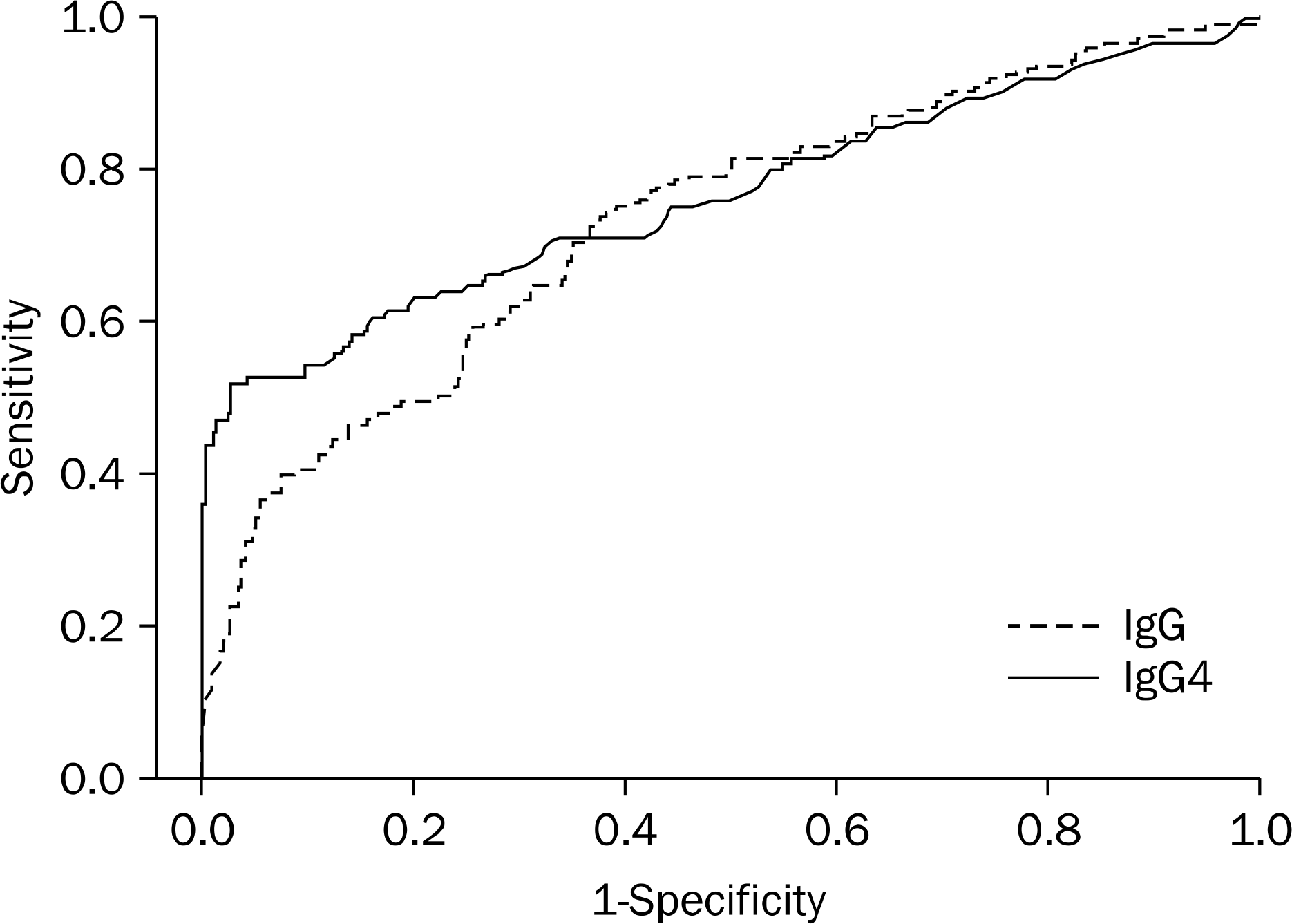 | Fig. 3.Receiver operating characteristics (ROC) curve of serum IgG and IgG4 in patients with autoimmune pancreatitis (AIP). The sensitivity and specificity of IgG4 at 133 mg/dL was 52% and 97%, respectively, in the differentiation of AIP from pancreatobiliary malignancies (area under the ROC curve [AUC], 0.77). The sensitivity and specificity of IgG at 1,335 mg/dL was 78.4% and 55.6%, respectively (AUC, 0.73). |
Table 1.
Korean Criteria for Autoimmune Pancreatitis (by Korean Pancreatobiliary Association, 2007)
Table 2.
Comparision of Patients with AIP and Pancreatobiliary Malignancies
Table 3.
Sensitivity and Specificity of IgG4 in the Diagnosis of AIP
Table 4.
The Elevation of Serum IgG and IgG4 in Patients with Autoimmune Pancreatitis (n=125)
| IgG4 elevation (>135 mg/dL) | ||
|---|---|---|
| Yes | No | |
| IgG elevation Yes | 34 (27.2) | 20 (16) |
| (≥1,800 mg/dL) No | 31 (24.8) | 40 (32) |
Table 5.
Serum CEA and CA 19–9 Levels of AIP and Pancreatobiliary Malignancies
 XML Download