Abstract
Background/Aims
Incidentally detected focal 18 F-fluorodeoxyglucose (FDG) uptake was compared with colonoscopy. We investigated the characteristics of colon adenomas which were revealed on PET/CT. Then we identified whether additional colonoscopy was necessary in patients with lesions which were revealed on PET/CT but had no matched lesions on colonoscopy.
Methods
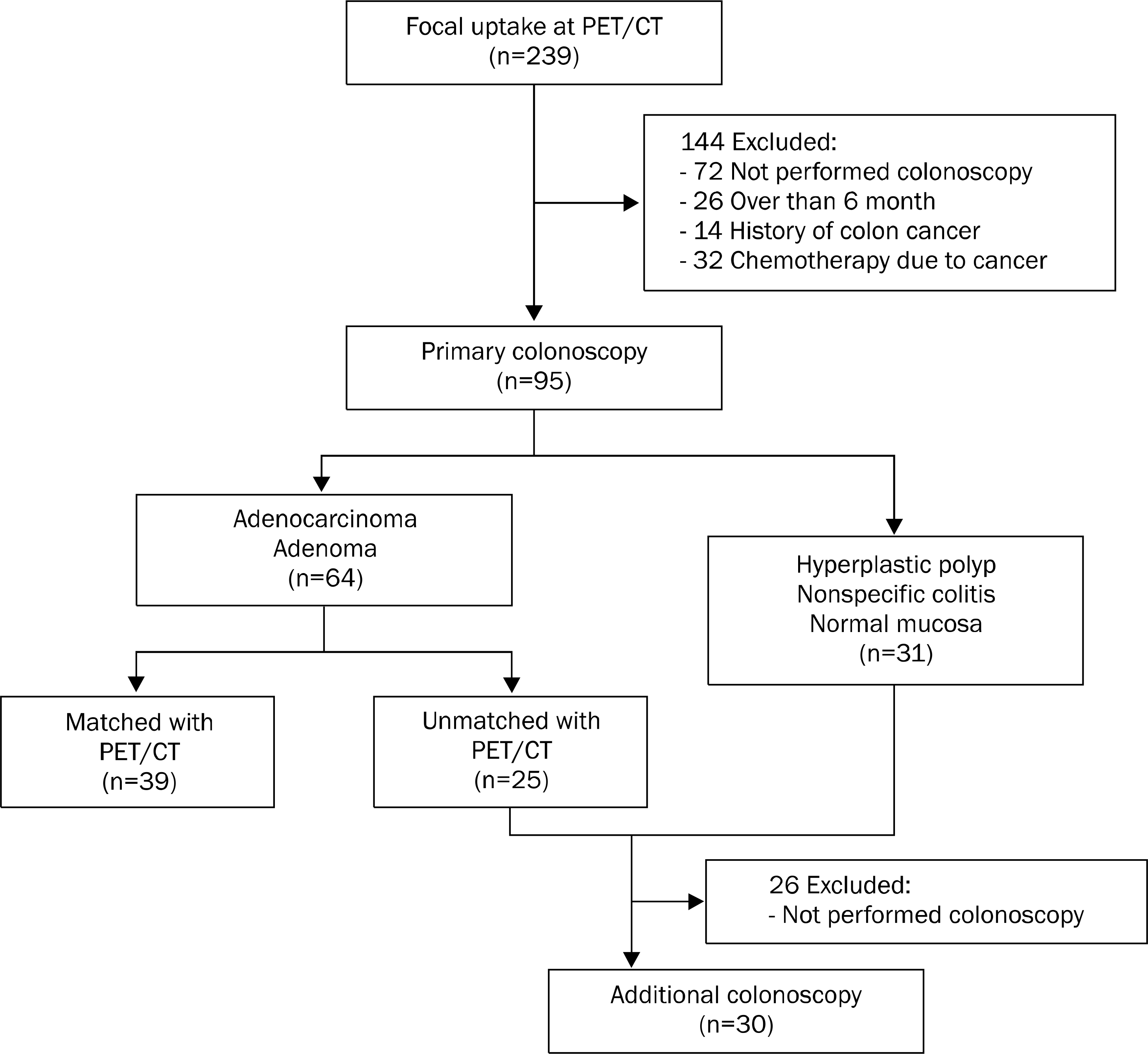
We retrospectively reviewed 95 patients who underwent colonoscopy within a 6 month interval after they had focal FDG uptake from January 2010 to May 2012 at National Police Hospital in Korea. Also, we analyzed 30 patients who underwent additional colonoscopy within 2 years after they had no matched lesions on primary colonoscopy.
Results
PET/CT depicted 54.6% (41/75) of adenomas and adenocarcinomas. The PET visibility of colon adenoma was significantly associated with degree of dysplasia (p=0.027), histologic type (p=0.040), and the size (p=0.038). The positivity rate was increased with higher degree of dysplasia (low-grade dysplasia, 47%; high-grade dysplasia, 78%; adenocarcinoma, 100%) and villous patterns of histologic type (tubular, 46.8%; tubulovillous, 87.5%; villous, 100%). Patients with adenomas larger than 10 mm (87.5%) had higher detection rate compared to those with adenomas smaller than 10 mm (49.0%). Among the 30 patients who underwent additional colonoscopy, only one patient had a 6 mm sized tubular adenoma (low-grade dysplasia).
Conclusions
Incidental focal colonic uptake may indicate advanced adenoma or adenocarcinoma. Thus, it justifies performing colonoscopy for identifying the presence of colon neoplasms. However, in case of unmatched lesions between PET/CT and colonoscopy, there was little evidence that additional colonoscopy would yield benefits.
Go to : 
References
1. Gallagher BM, Fowler JS, Gutterson NI, MacGregor RR, Wan CN, Wolf AP. Metabolic trapping as a principle of oradiophar-maceutical design: some factors resposible for the biodistri-bution of [18F] 2-deoxy-2-fluoro-D-glucose. J Nucl Med. 1978; 19:1154–1161.
2. Gambhir SS, Czernin J, Schwimmer J, Silverman DH, Coleman RE, Phelps ME. A tabulated summary of the FDG PET literature. J Nucl Med. 2001; 42(5 Suppl):1S–93S.
3. Schöder H, Gönen M. Screening for cancer with PET and PET/CT: potential and limitations. J Nucl Med. 2007; 48(Suppl 1):4S–18S.
4. Shin SJ, Choi JW, Lee SK, Choi CH, Kim TI, Kim WH. A case of multiple colonic adenomas which were found incidentally in FDG-PET. Korean J Med. 2004; 66:639–643.
5. Patrikeos AP, Mackay JR, Hicks RJ. Detection of synchronous adenocarcinomas and multiple dysplastic polyps with F-18 FDG positron emission tomography in a case of nonfamilial polyposis. Clin Nucl Med. 2003; 28:487–488.

6. Okuno T, Fu KI, Sano Y, et al. Early colon cancers detected by FDG-pet: a report of two cases with immunohistochemical investigation. Hepatogastroenterology. 2004; 51:1323–1325.
7. Yasuda S, Fujii H, Nakahara T, et al. 18F-FDG PET detection of colonic adenomas. J Nucl Med. 2001; 42:989–992.
8. Gutman F, Alberini JL, Wartski M, et al. Incidental colonic focal lesions detected by FDG PET/CT. AJR Am J Roentgenol. 2005; 185:495–500.

9. van Kouwen MC, Nagengast FM, Jansen JB, Oyen WJ, Drenth JP. 2-(18F)-fluoro-2-deoxy-D-glucose positron emission tomography detects clinical relevant adenomas of the colon: a prospective study. J Clin Oncol. 2005; 23:3713–3717.

10. Chen YK, Kao CH, Liao AC, Shen YY, Su CT. Colorectal cancer screening in asymptomatic adults: the role of FDG PET scan. Anticancer Res. 2003; 23:4357–4361.
11. Nakajo M, Jinnouchi S, Tashiro Y, et al. Effect of clinicopathologic factors on visibility of colorectal polyps with FDG PET. AJR Am J Roentgenol. 2009; 192:754–760.

12. Hixson LJ, Fennerty MB, Sampliner RE, McGee D, Garewal H. Prospective study of the frequency and size distribution of polyps missed by colonoscopy. J Natl Cancer Inst. 1990; 82:1769–1772.

13. Bensen S, Mott LA, Dain B, Rothstein R, Baron J. The colonoscopic miss rate and true one-year recurrence of colorectal neoplastic polyps. Polyp Prevention Study Group. Am J Gastroenterol. 1999; 94:194–199.
14. Rex DK, Cutler CS, Lemmel GT, et al. Colonoscopic miss rates of adenomas determined by back-to-back colonoscopies. Gastroenterology. 1997; 112:24–28.

15. Kasugai K, Miyata M, Hashimoto T, et al. Assessment of miss and incidence rates of neoplastic polyps at colonoscopy. Dig Endosc. 2005; 17:44–49.

16. Drenth JP, Nagengast FM, Oyen WJ. Evaluation of (pre-)malignant colonic abnormalities: endoscopic validation of FDG-PET findings. Eur J Nucl Med. 2001; 28:1766–1769.

17. Schlemper RJ, Riddell RH, Kato Y, et al. The Vienna classification of gastrointestinal epithelial neoplasia. Gut. 2000; 47:251–255.

18. Brenner H, Hoffmeister M, Stegmaier C, Brenner G, Altenhofen L, Haug U. Risk of progression of advanced adenomas to colorectal cancer by age and sex: estimates based on 840,149 screening colonoscopies. Gut. 2007; 56:1585–1589.
19. Ono K, Ochiai R, Yoshida T, et al. The detection rates and tumor clinical/pathological stages of whole-body FDG-PET cancer screening. Ann Nucl Med. 2007; 21:65–72.

20. Yasunaga H. Who wants cancer screening with PET? A con-tingent valuation survey in Japan. Eur J Radiol. 2009; 70:190–194.
21. Lee JC, Hartnett GF, Hughes BG, Ravi Kumar AS. The segmental distribution and clinical significance of colorectal fluorodeoxyglucose uptake incidentally detected on PET-CT. Nucl Med Commun. 2009; 30:333–337.

22. Prabhakar HB, Sahani DV, Fischman AJ, Mueller PR, Blake MA. Bowel hot spots at PET-CT. Radiographics. 2007; 27:145–159.

23. Ahmad Sarji S. Physiological uptake in FDG PET simulating disease. Biomed Imaging Interv J. 2006; 2:e59.

24. Tatlidil R, Jadvar H, Bading JR, Conti PS. Incidental colonic fluorodeoxyglucose uptake: correlation with colonoscopic and histopathologic findings. Radiology. 2002; 224:783–787.

25. Kang MK, Hong SP, Lee JE, et al. The usefulness of F18-FDG PET/CT in detection of colonic neoplasm. Intest Res. 2010; 8:18–23.

26. Roh SH, Jung SA, Kim SE, et al. The clinical meaning of benign colon uptake in (18)F-FDG PET: comparison with colonoscopic findings. Clin Endosc. 2012; 45:145–150.
27. Pai M, Cho YK, Jung SA, Shim KN, Lee HS. Colonic uptake patterns of F-18-FDG PET in asymptomatic adults: comparison with colonoscopic findings. Korean J Nucl Med. 2005; 39:15–20.
28. Israel O, Yefremov N, Bar-Shalom R, et al. PET/CT detection of unexpected gastrointestinal foci of 18F-FDG uptake: incidence, localization patterns, and clinical significance. J Nucl Med. 2005; 46:758–762.
29. Delbeke D, Martin WH. PET and PET-CT for evaluation of colorectal carcinoma. Semin Nucl Med. 2004; 34:209–223.

30. Zhuang H, Yu JQ, Alavi A. Applications of fluorodeoxyglucose-PET imaging in the detection of infection and inflammation and other benign disorders. Radiol Clin North Am. 2005; 43:121–134.

31. Heresbach D, Barrioz T, Lapalus MG, et al. Miss rate for colorectal neoplastic polyps: a prospective multicenter study of back-to-back video colonoscopies. Endoscopy. 2008; 40:284–290.

32. Farrar WD, Sawhney MS, Nelson DB, Lederle FA, Bond JH. Colorectal cancers found after a complete colonoscopy. Clin Gastroenterol Hepatol. 2006; 4:1259–1264.

33. Weston AP, Campbell DR. Diminutive colonic polyps: histopathology, spatial distribution, concomitant significant lesions, and treatment complications. Am J Gastroenterol. 1995; 90:24–28.
34. Macrae FA, Tan KG, Williams CB. Towards safer colonoscopy: a report on the complications of 5000 diagnostic or therapeutic colonoscopies. Gut. 1983; 24:376–383.

35. Nivatvongs S. Complications in colonoscopic polypectomy. An experience with 1,555 polypectomies. Dis Colon Rectum. 1986; 29:825–830.
36. Rosen L, Bub DS, Reed JF 3rd, Nastasee SA. Hemorrhage following colonoscopic polypectomy. Dis Colon Rectum. 1993; 36:1126–1131.

Go to : 
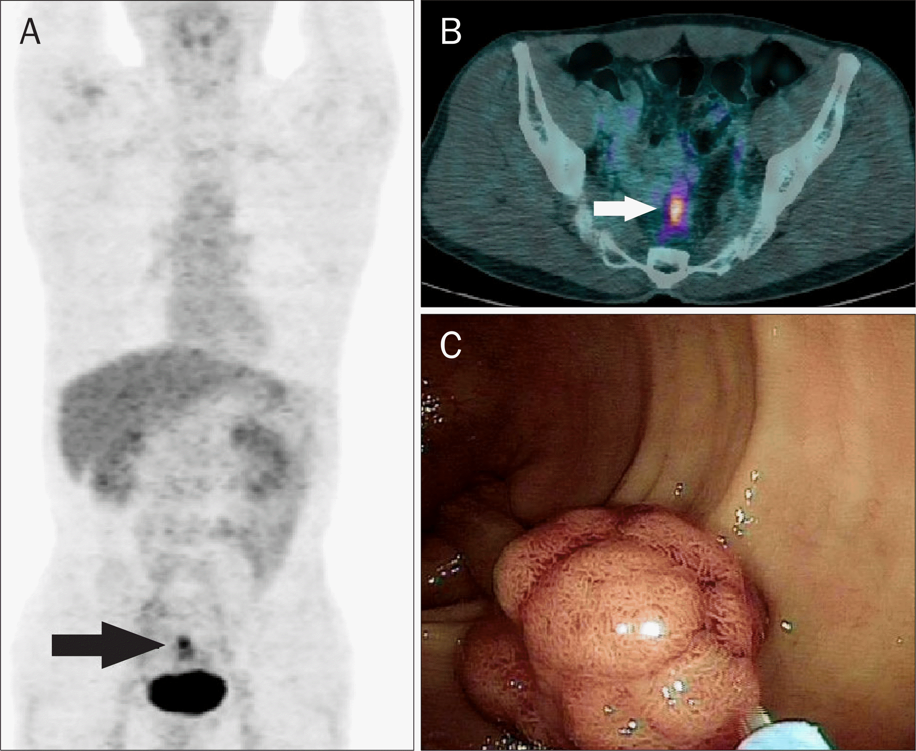
 | Fig. 2.PET (A) and PET/CT (B) images showed an focal increased 18 F-fluoro-deoxyglucose uptake (A, black arrow; B, white arrow) at the sigmoid colon in a 51-year-old asymptomatic man. (C) A 20-mm sized adenocarcinoma of the sigmoid colon was observed and removed with colonoscopy. |
Table 1.
Comparison of PET/CT and Colonoscopic Results
| FDG uptake in PET/CT | Total | ||
|---|---|---|---|
| Focal uptake | No uptake | ||
| Colonoscopy | |||
| Positive Negativea | 4158 | 34 | 75 58 |
| Total | 99 | 34 | 133 |
Table 2.
Comparison of PET/CT Visibility of Adenoma according to the Histology


 PDF
PDF ePub
ePub Citation
 Print
Print


 XML Download