Abstract
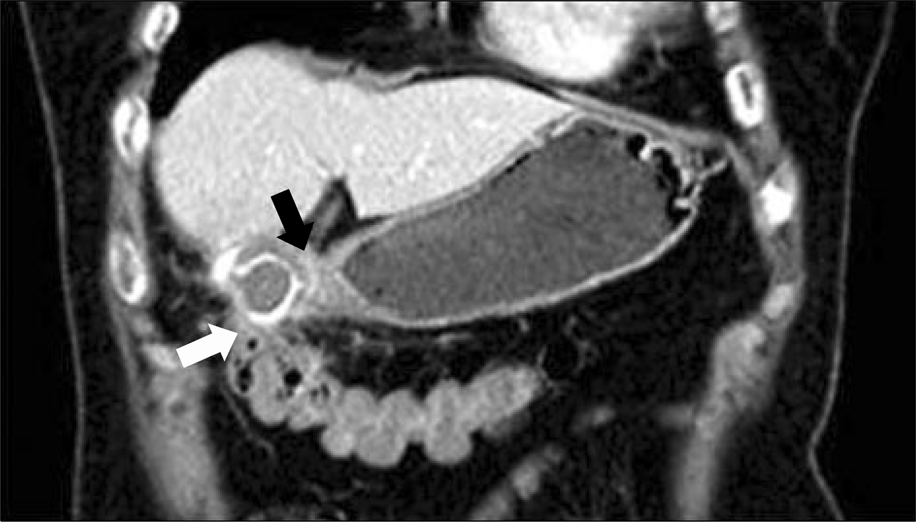
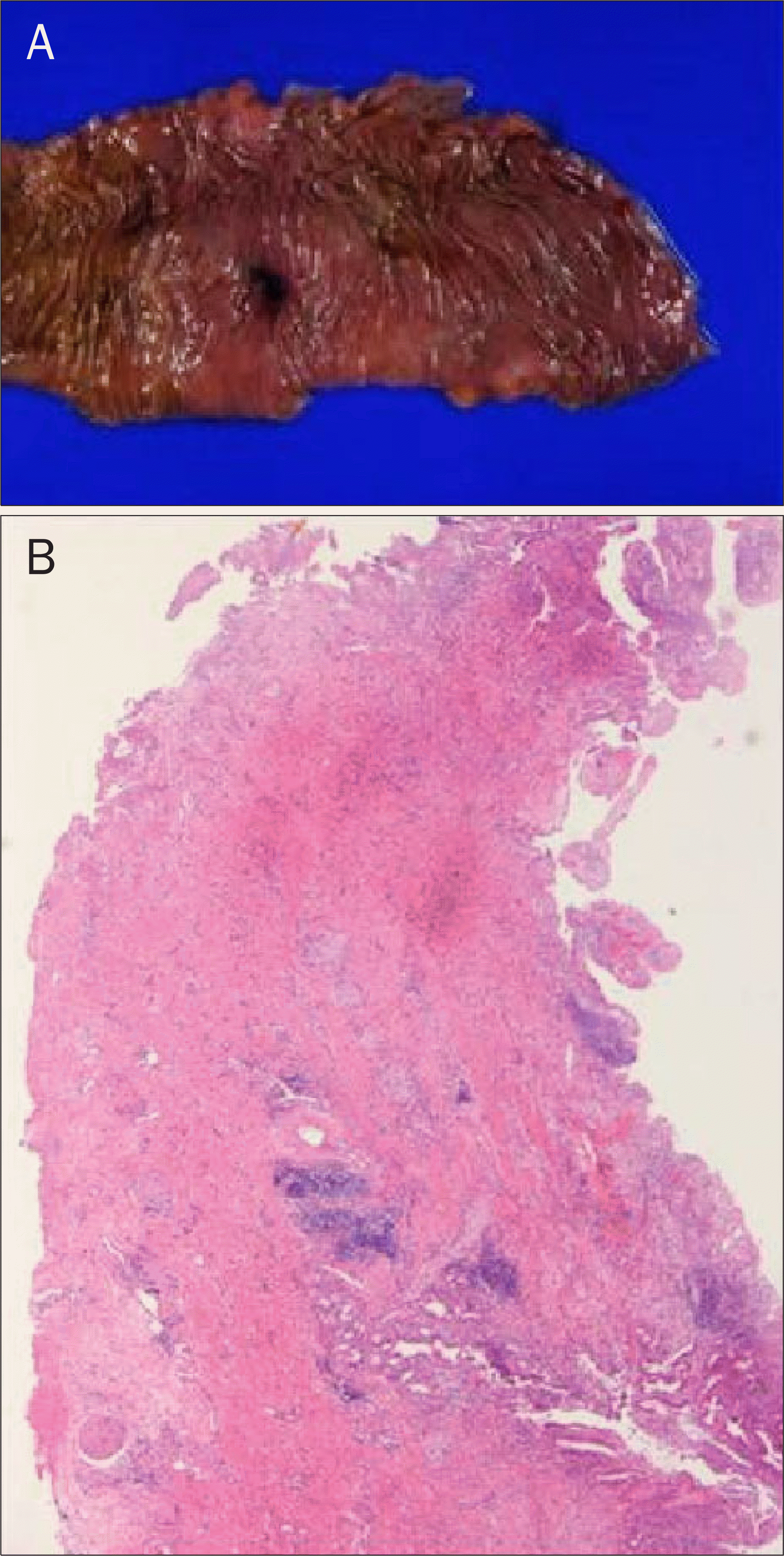
Biliary enteric fistula is an abnormal pathway often caused by biliary disease. It is difficult to diagnose the disease because patients have nonspecific symptoms. A 67-year-old woman presented with hematemesis and melena. She was diagnosed with Dieulafoy lesion on the gastric antrum and underwent endoscopic hemostasis using hemoclips. Follow-up upper gastrointestinal endoscopy revealed an abnormal opening on a previous treated site that was suggestive of biliary enteric fistula. Abdomen simple X-ray and abdominal dynamic CT scan showed pneumobilia and cholecysto-gastric fistula. The patient had cholecystectomy and wedge resection of the gastric antrum, followed by right extended hemicolectomy because of severe adhesive lesion between the gallbladder and colon. She was diagnosed with cholecysto-gastro-colic fistula postoperatively. We report on this case and give a brief review of the literatures.
References
1. Shim CS, Lee JS, Lee MS, et al. Cholecysto – duodeno – colic fistula: report of one case. Korean J Gastrointest Endosc. 1996; 16:801–806.
2. Yamashita H, Chijiiwa K, Ogawa Y, Kuroki S, Tanaka M. The internal biliary fistula–reappraisal of incidence, type, diagnosis and management of 33 consecutive cases. HPB Surg. 1997; 10:143–147.
3. Glenn F, Reed C, Grafe WR. Biliary enteric fistula. Surg Gynecol Obstet. 1981; 153:527–531.
4. Jang LC, Han SH, Kim SW, Park YH. Biliary-enteric fistula. Korean J Gastroenterol. 1992; 24:827–832.
5. Seo JS, Yang SY, Kim JH, et al. A case of choledocho-duode-no-colonic fistula. Korean J Gastrointest Endosc. 2007; 34:278–281.
6. Hakim M, Boyd R, Stricoff R, Shaftan G, Saxe A. Cholecystogas-trocolonic fistula with intrahepatic abscess: a rare complication of biliary stone disease. Am Surg. 1997; 63:472–474.
7. Elsas LJ, Gilat T. Cholecystocolonic fistula with malabsorption. Ann Intern Med. 1965; 63:481–486.

8. LeBlanc KA, Barr LH, Rush BM. Spontaneous biliary enteric fistulas. South Med J. 1983; 76:1249–1252.

9. La Greca G, Grasso E, Sofia M, Gagliardo S, Barbagallo F. Complicated duodeno-biliary fistula in bleeding duodenal ulcer: case report an literature review. Ann Ital Chir. 2008; 79:57–61.
10. Stempfle B, Diamantopoulos G. Spontaneous cholecysto-ga-stric fistula with massive gastrointestinal bleeding. Endoscopic diagnosis and concrement extraction. Fortschr Med. 1976; 94:444–447.
11. Feller ER, Warshaw AL, Schapiro RH. Observations on management of choledochoduodenal fistula due to penetrating peptic ulcer. Gastroenterology. 1980; 78:126–131.

12. Miyamoto S, Furuse J, Maru Y, Tajiri H, Muto M, Yoshino M. Duodenal tuberculosis with a choledochoduodenal fistula. J Gastroenterol Hepatol. 2001; 16:235–238.

13. Rutgeerts P, Onette E, Vantrappen G, Geboes K, Broeckaert L, Talloen L. Crohn's disease of the stomach and duodenum: A clinical study with emphasis on the value of endoscopy and endoscopic biopsies. Endoscopy. 1980; 12:288–294.

14. Min SK, Han HS, Kim EG, Kim YW, Choi YM. Laparoscopic repair of biliary-enteric fistula – report of 3 cases. J Korean Soc Endosc Laparosc Surg. 2002; 5:20–24.
15. LeBlanc KA. General laparoscopic surgical complications. LeBlanc KA, editor. Management of laparoscopic surgical complications. New York: Marcel Dekker;2004. p. 43–62.
Fig. 1.
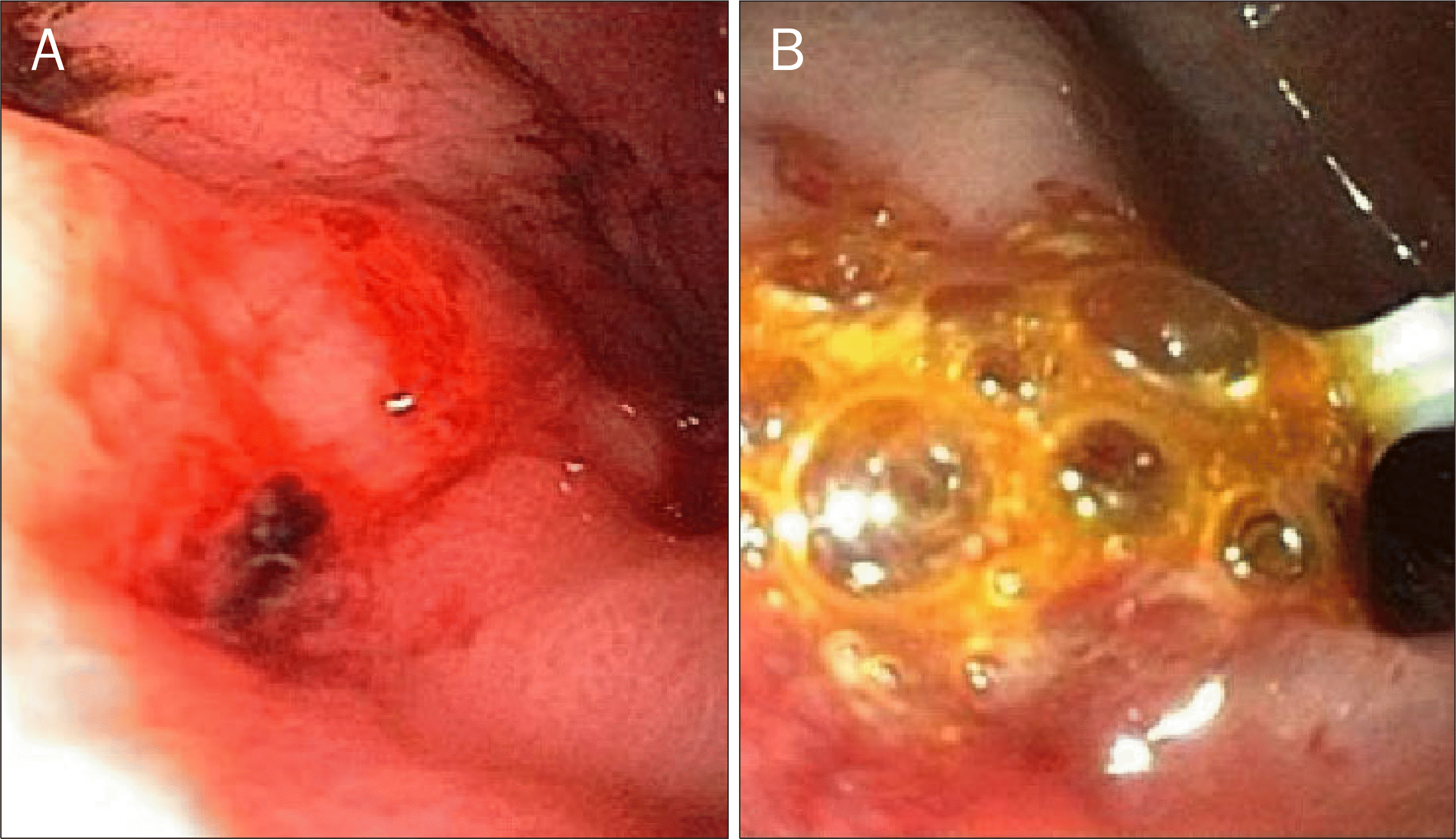
Upper gastrointestinal endoscopic findings. (A) Fresh blood from an exposed vessel was observed on the great curvature of the gastric antrum. (B) Follow-up examination showed an abnormal opening at the gastric antrum. Yellowish and bubbly contents were drained through the lesion.

Fig. 2.
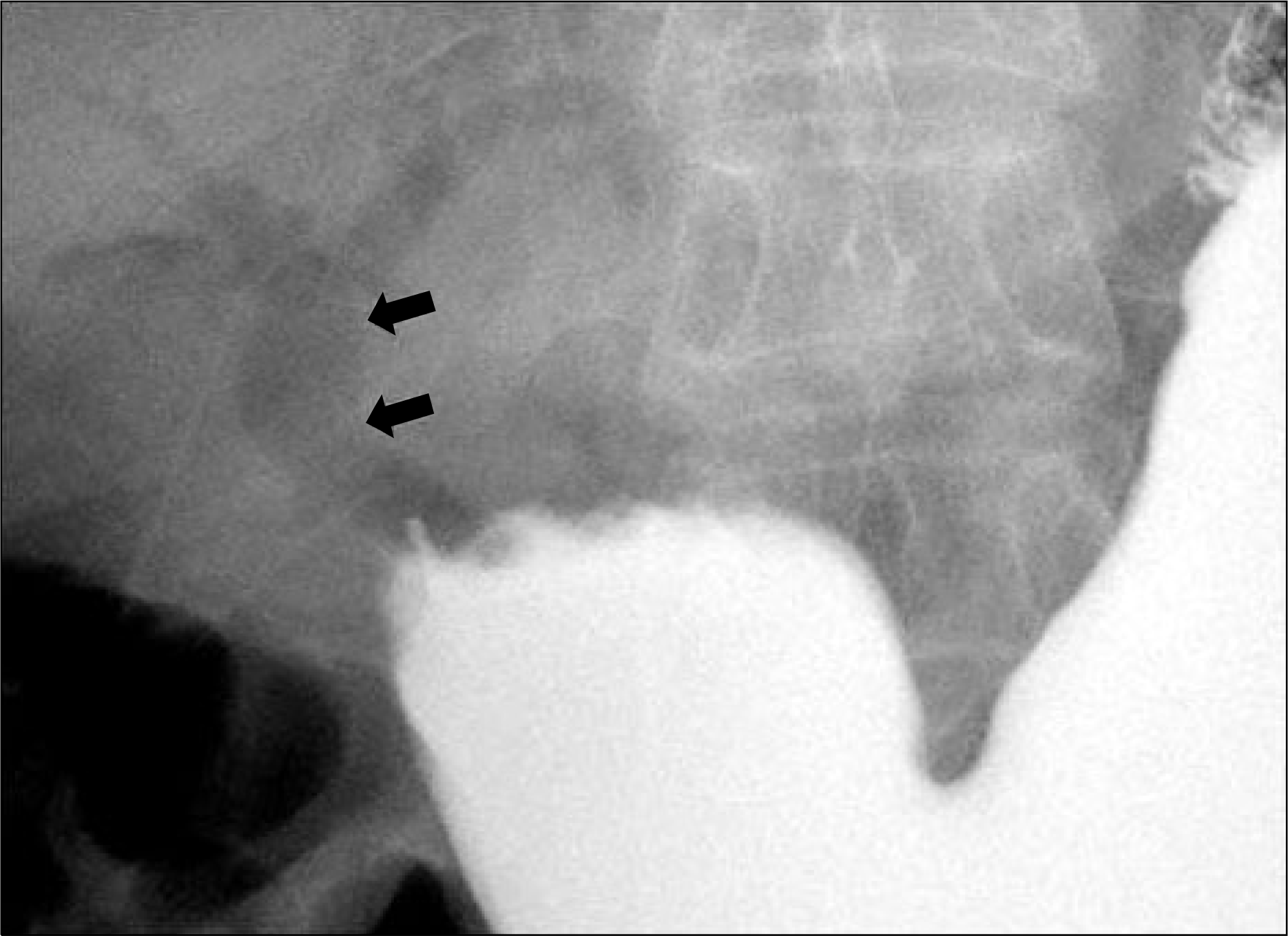
Abdomen simple X-ray. It revealed pneumobilia (arrows) in the common bile duct, suggesting the presence of biliary enteric fistula.


 PDF
PDF ePub
ePub Citation
 Print
Print


 XML Download