

 PDF
PDF ePub
ePub Citation
 Print
Print


Abstract
Intestinal malrotation is a congenital disorder that results from the failure of normal bowel rotation and fixation during the 5th gestational week. The incidence of intestinal malrotation is <0.2%, but prompt diagnosis is important because this anomaly can cause midgut volvulus and lead to fatalities. Compared to infants presenting with acute symptoms, such as abdominal pain, vomiting, or diarrhea, adult patients complain of intermittent self-limited abdominal pain. We present a case of intestinal malrotation complicated by midgut volvulus improved with conservative care in a 70-year-old man. The diagnosis was suggested on the basis of imaging findings.
References
1. Sheridan M. Nonrotation of the midgut presenting in the adolescent and adult. Am J Gastroenterol. 1989; 84:670–673.
2. Rowsom JT, Sullivan SN, Girvan DP. Midgut volvulus in the adult. A complication of intestinal malrotation. J Clin Gastroenterol. 1987; 9:212–216.
3. Firor HV, Harris VJ. Rotational abnormalities of the gut. Re-emphasis of a neglected facet, isolated incomplete rotation of the duodenum. Am J Roentgenol Radium Ther Nucl Med. 1974; 120:315–321.
4. Kanazawa T, Kasugai K, Miyata M, et al. Midgut malrotation in adulthood. Intern Med. 2000; 39:626–631.
5. Emanuwa OF, Ayantunde AA, Davies TW. Midgut malrotation first presenting as acute bowel obstruction in adulthood: a case report and literature review. World J Emerg Surg. 2011; 6:22.

6. Stewart DR, Colodny AL, Daggett WC. Malrotation of the bowel in infants and children: a 15 year review. Surgery. 1976; 79:716–720.
7. Ford EG, Senac MO Jr, Srikanth MS, Weitzman JJ. Malrotation of the intestine in children. Ann Surg. 1992; 215:172–178.
8. Cho YH, Kim HY. A clinical review of symptomatic intestinal malrotation. J Korean Surg Soc. 2007; 73:246–249.
9. von Flüe M, Herzog U, Ackermann C, Tondelli P, Harder F. Acute and chronic presentation of intestinal nonrotation in adults. Dis Colon Rectum. 1994; 37:192–198.
10. Shimanuki Y, Aihara T, Takano H, et al. Clockwise whirlpool sign at color Doppler US: an objective and definite sign of midgut volvulus. Radiology. 1996; 199:261–264.
11. Leonidas JC, Magid N, Soberman N, Glass TS. Midgut volvulus in infants: diagnosis with US. Work in progress. Radiology. 1991; 179:491–493.
12. Sizemore AW, Rabbani KZ, Ladd A, Applegate KE. Diagnostic performance of the upper gastrointestinal series in the evaluation of children with clinically suspected malrotation. Pediatr Radiol. 2008; 38:518–528.
13. El-Gohary Y, Alagtal M, Gillick J. Longterm complications following operative intervention for intestinal malrotation: a 10-year review. Pediatr Surg Int. 2010; 26:203–206.
14. Choi M, Borenstein SH, Hornberger L, Langer JC. Heterotaxia syndrome: the role of screening for intestinal rotation abnormalities. Arch Dis Child. 2005; 90:813–815.
15. Nehra D, Goldstein AM. Intestinal malrotation: varied clinical presentation from infancy through adulthood. Surgery. 2011; 149:386–393.
16. Choi JW, Kim KH, Seo HJ, et al. A case of intestinal nonrotation incidentally detected on DISIDA scan. Korean J Med. 2002; 62:566–569.
17. Park HN, Park JJ, Cheon JH, et al. Nonrotation of the prearterial segment of midgut presenting as duodenal obstruction in a 60-year-old man. Korean J Gastroenterol. 2010; 55:252–255.
Fig. 1.
(A) Abdominal CT demonstrated the superior mesenteric artery (SMV) (arrow) located in the upper abdomen and to the left of the SMA (arrow head), inversion of the SMA and superior mesenteric vein. (B) Whirling appearance of mesenteric vessels around the SMA.

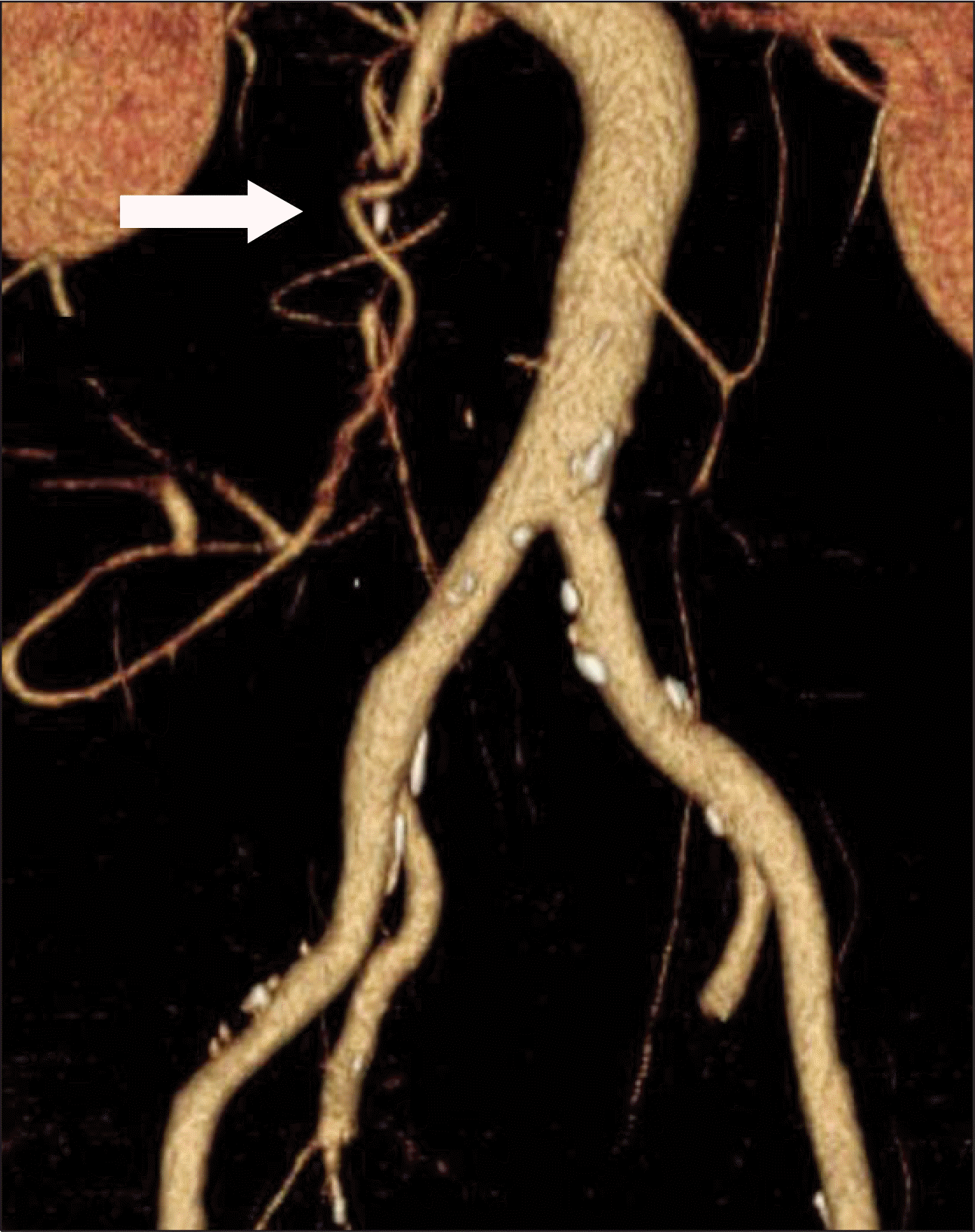
Fig. 2.
CT angiography showed clockwise corkscrew appearance (barber's pole sign) of the superior mesenteric vein (arrow).
 XML Download