

 PDF
PDF ePub
ePub Citation
 Print
Print


Abstract
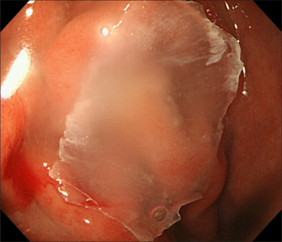
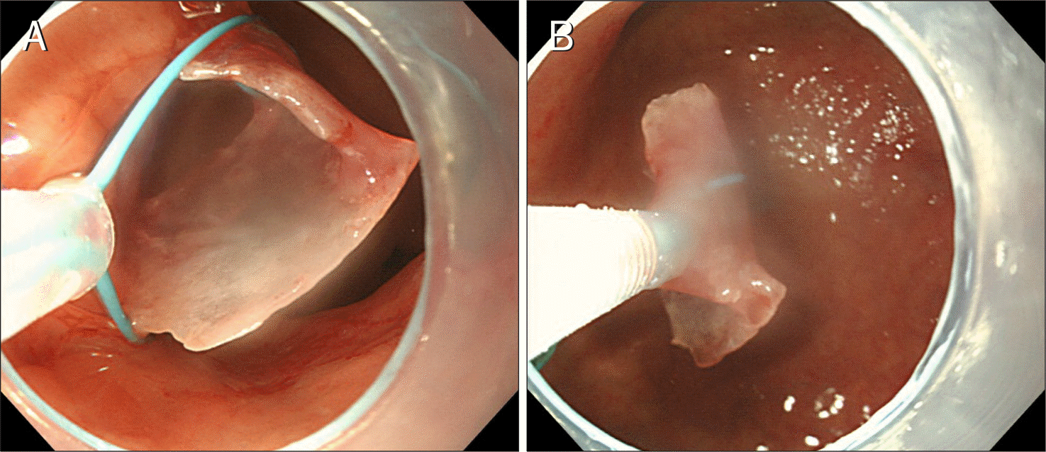
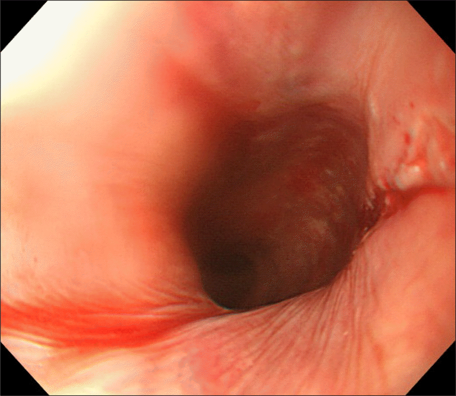
A sharp, impacted fish bone in the esophagus is an indication for urgent endoscopy. Endoscopic removal of such an object is a challenging task. An endoscopic protector hood is then used to remove the object. However, an endoscopic hood protector is not always available. In a patient with a large hiatal hernia, the protector hood may not return to the original shape when it passes through the gastroesophageal junction and therefore may not properly protect the esophageal mucosa from the sharp foreign body. In our case, it was impossible to deploy the endoscopic hood protector through the gastroesophageal junction despite multiple attempts. We propose an alternative solution for such cases. We safely removed a large sharp-edged flat fish bone that was folded and compressed using a detachable snare after releasing and pushing the fish bone into the stomach using an endoscope equipped with a transparent cap used for dilating the esophageal wall. This method of using an endoscopic cap and detachable snare is a safe, useful alternative for endoscopically removing a large sharp-edged flat foreign body from the upper gastrointestinal tract. This alternative technique has not been reported in the English medical literature.
References
1. Bertoni G, Sassatelli R, Conigliaro R, Bedogni G. A simple latex protector hood for safe endoscopic removal of sharp-pointed gastroesophageal foreign bodies. Gastrointest Endosc. 1996; 44:458–461.

2. Jeen YT, Chun HJ, Song CW, et al. Endoscopic removal of sharp foreign bodies impacted in the esophagus. Endoscopy. 2001; 33:518–522.
3. Brady PG. Esophageal foreign bodies. Gastroenterol Clin North Am. 1991; 20:691–701.
4. Sittitrai P, Pattarasakulchai T, Tapatiwong H. Esophageal foreign bodies. J Med Assoc Thai. 2000; 83:1514–1518.
5. Ashraf O. Foreign body in the esophagus: a review. Sao Paulo Med J. 2006; 124:346–349.
6. Li ZS, Sun ZX, Zou DW, Xu GM, Wu RP, Liao Z. Endoscopic management of foreign bodies in the upper-GI tract: experience with 1088 cases in China. Gastrointest Endosc. 2006; 64:485–492.
7. Higo R, Matsumoto Y, Ichimura K, Kaga K. Foreign bodies in the aerodigestive tract in pediatric patients. Auris Nasus Larynx. 2003; 30:397–401.
8. Vizcarrondo FJ, Brady PG, Nord HJ. Foreign bodies of the upper gastrointestinal tract. Gastrointest Endosc. 1983; 29:208–210.
9. Eisen GM, Baron TH, Dominitz JA, et al. American Society for Gastrointestinal Endoscopy. Guideline for the management of ingested foreign bodies. Gastrointest Endosc. 2002; 55:802–806.
10. Katsinelos P, Kountouras J, Paroutoglou G, Zavos C, Mimidis K, Chatzimavroudis G. Endoscopic techniques and management of foreign body ingestion and food bolus impaction in the upper gastrointestinal tract: a retrospective analysis of 139 cases. J Clin Gastroenterol. 2006; 40:784–789.
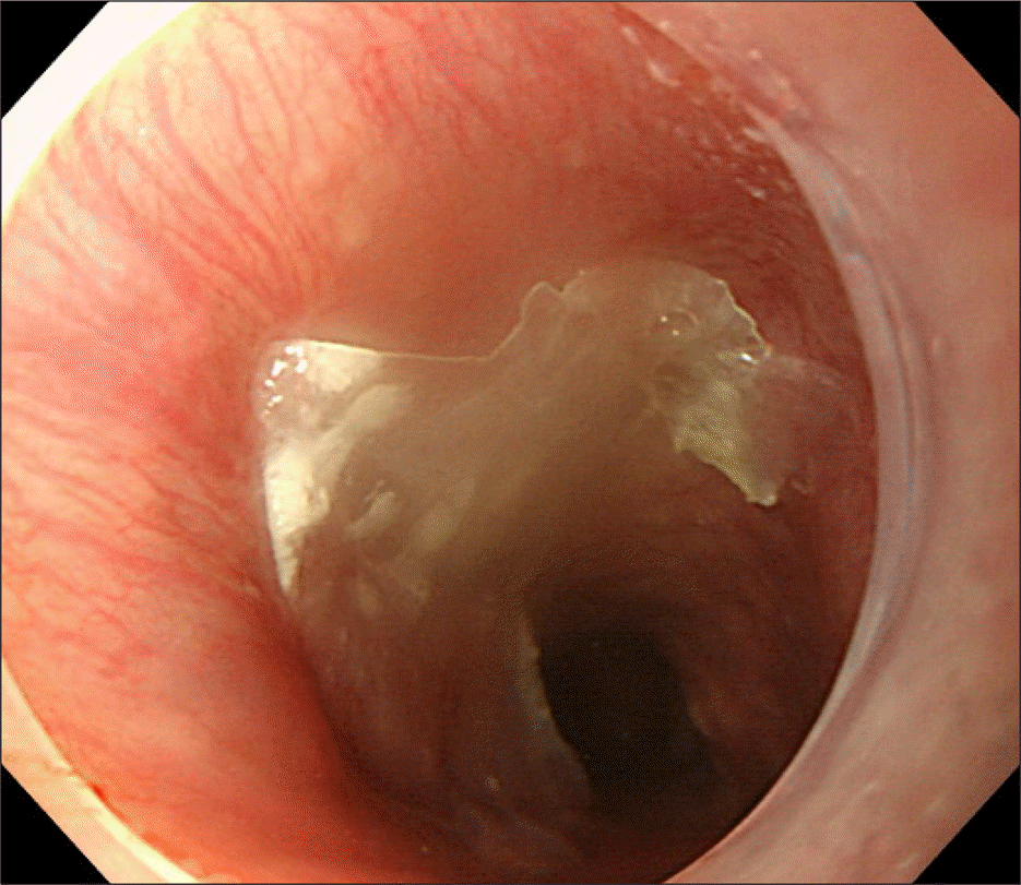
Fig. 1.
The endoscope revealed that a large sharp-edged flat fish bone impacted just below the upper esophageal sphincter, 25 cm from the incisors.
 XML Download