

 PDF
PDF ePub
ePub Citation
 Print
Print


INTRODUCTION
Heterotaxy is a rare condition wherein the orderly arrangement of organs and blood vessels, as seen in situs solitus and situs inversus, is lost.1 In addition, it has multiple congenital anomalies. Polysplenia is a subtype of heterotaxy (asplenia is another) characterized by cardiac malformations (both atria displaying the morphology of the left atrium), bilobed lung (each lung having the morphology of a left lung), visceral malrotation, multiple spleens, interruption of the inferior vena cava with azygos or hemizygos continuation, and preduodenal portal vein.2,3 Patients with polysplenia sometimes have short pancreas or agenesis of dorsal pancreas, which is related to the possibility of increased incidence of pancreatitis.4,5
EUS provides high-resolution images of the entire pancreatic parenchyma and allows direct visualization of the ductal system.6 Therefore, EUS may be helpful in the diagnosis of polysplenia syndrome with agenesis of the dorsal pancreas when CT scans and MRI were inconclusive for the pancreatic lesion. Herein, we report a case of agenesis of the dorsal pancreas in association with polysplenia syndrome that manifested as acute pancreatitis.
CASE REPORT
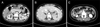
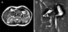
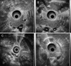
A 49-year-old female was referred to our hospital for acute persistent epigastric pain for 1 week prior to admission. She had no other illnesses and no remarkable family medical history. She denied any history of drug or alcohol abuse. The laboratory findings were normal except for elevated of serum amylase (2,023 IU/L, normal range 36-128 IU/L) and lipase (1,312 U/L, normal range 22-51 U/L) levels. Therefore, she was diagnosed with acute pancreatitis. Additional examinations were done to find the cause of pancreatitis. The serum calcium level was 8.6 mg/dL and triglyceride level was 52 mg/dL. The serum IgG4 level was 25 mg/dL. Abdomen CT scan revealed a midline liver with hepatomegaly, a centrally located gallbladder, multiple spleens in the left upper quadrant behind the stomach, an inferior vena cava interruption with an azygos continuation, preduodenal portal vein, and a right-sided small bowel and left-sided large bowel (Fig. 1). A short pancreas with a prominent head and no tail was evident. MRI demonstrated that main pancreatic duct was short and the accessory duct was absent (Fig. 2). EUS revealed a hypertrophied pancreatic head with a main pancreatic duct of normal caliber, absence of the accessory pancreatic duct, homogenous echogenicity throughout the pancreatic parenchyme, splenic vessels that were contact with the posterior wall of the stomach, and a preduodenal portal vein crossing over the pancreatic head. In addition, there were no sludges or stones in the gallbladder and bile duct and no pancreatic divisum (Fig. 3). Endoscopy revealed a normal major papilla but the absence of the minor papilla. All these findings led to the diagnosis of dorsal pancreatic agenesis with polysplenia syndrome. After fasting and conservative treatment, the patient's symptoms improved, and her serum amylase and lipase levels were normalized. No evidence of relapse was noted in the 12-month follow-up.
Discussion
Polysplenia syndrome is a rare anomaly that occurs in approximately 4 of every million live births.7 It is complex set of various visceral anomalies including multiple spleens, impaired visceral lateralization, cardiovascular anomalies, gastrointestinal abnormalities, and short pancreas.1 Rarely, genitourinary anomalies such as double ureters, renal agenesis or hypoplastic kidney are reported as part of the polysplenia syndrome.8 However, this syndrome does not have fixed set of characteristics that are present in all cases.1
The unique sign of the polysplenia syndrome is the presence of multiple spleens, mostly located throughout the greater curvature of the stomach, which is to the right of the midline in more than 60% of patients.9
Cardiac anomaly is frequently associated with polysplenia syndromes in children but this is far less frequent in adults because of early decease in children with this anomaly.2 Cardiovascular anomalies associated with polysplenia syndrome include the absence or hypoplasia of the suprarenal inferior vena cava (with or without azygos or hemiazygos continuation), dextrocardia, ventricular septal defects, and morphologic left ventricular outflow obstruction, and these anomalies lead patients to death with congestive heart failure, hypoplastic left ventricle or morbidity of cardiac operation before 5 years old.2 Our patient did not have specific past history about heart disease, in addition, electrocardiography and echocardiography was normal.
Preduodenal portal vein is a portal vein located in front of the pancreatic head at CT image. It passes ventral to the duodenum and the head of the pancreas. Preduodenal portal vein might interfere mechanically with pancreatic development, so it is associated with annular pancreas and malrotation.3 This vascular anomaly has importance in points of the prevention of accident during pancreaticobiliary operation.10
Abdominal complaints are the most common symptoms in adult, and sometimes CT scan shows intestinal malrotation. It is usually an incidental finding in polysplenia but occasionally can cause abdominal pain via recurrent volvulus or obstruction from a mesenteric band.11 In some cases, pancreatitis with complete agenesis of dorsal pancreas can cause abdominal pain.5 Sempere et al.12 reviewed 14 cases of complete agenesis of dorsal pancreas and reported abdominal pain (13 cases, 92.9%) and pancreatitis (7 cases, 50%). Rakesh et al.5 showed that 4 cases with complete agenesis of dorsal pancreas had pancreatitis and abdominal pain. In complete agenesis of dorsal pancreas, pancreatitis may be related with sphincter of Oddi dysfunction or higher intrapancreatic duct pressures due to hypertrophy of the ventral gland.13,14 In our cases, there were no other causes of pancreatitis. However, hyperplasia of ventral pancreas was noted at EUS and might cause increased intrapancreatic duct pressure or hypersecretion of pancreatic enzyme resulting in pancreatitis.
Embryologically, the pancreas arises from ventral and dorsal buds. While ventral bud forms the uncinate process and pancreatic head, the dorsal bud becomes the body and tail. The dorsal pancreas and spleen develop in the dorsal mesogastrium. Therefore, concomitant anomalies of both organs can be expected in patient with polysplenia syndrome.4 Short pancreas in polysplenia syndrome means partial agenesis or hypoplasia of the dorsal pancreas.15 In partial agenesis, the minor papilla with a remnant of the accessory pancreatic duct and the body of the pancreas are present but the minor papilla, accessory pancreatic duct, body and tail of the pancreas are absent in complete agenesis.15,16 Partial agenesis of the pancreas can remain asymptomatic because of the functional reserves of the exocrine and the endocrine pancreas.17 The clinical significance in complete agenesis is a possible relationship to early or late onset diabetes mellitus, probably because most of the islet cells are located in the pancreatic body and tail.18,19 However, our patient did not have diabetes and fasting sugar was 99 mg/dL.
We confirmed polysplenia syndrome with CT scans and MRI/MRCP but confused whether she had partial agenesis of dorsal pancreas or complete agenesis of dorsal pancreas with hypertrophied ventral pancreas. ERCP is considered the gold standard for the diagnosis of agenesis of the dorsal pancreas.16 However, ERCP is an invasive method, and it is also technically difficult to identify the minor papilla and to cannulate a catheter into it.16 CT scans, MRCP and ERCP have some limit in distinguishing agenesis of the dorsal pancreas from other congenital abnormalities (pancreatic divisum, annular pancreas, etc.).4,12 Sempere et al.12 showed that EUS was helpful in the identification of agenesis of the dorsal pancreas when other imaging modalities were inconclusive. EUS can directly visualize the body and tail of the pancreas and preduodenal portal vein during the retraction the duodenum to the gastric antrum (called as "stripping").12 With the EUS positioned in the gastric antrum, the body and the tail of the pancreas are visualized between the posterior wall of the stomach and the splenic vessels.6 Agenesis of the dorsal pancreas may be considered, as in our patient, if the accessory pancreatic duct, and the body and tail of pancreas (without any structure between the stomach and the splenic vessels) are not visualized.12 If the pancreatic parenchyma showed homogenous echogenicity during stripping, we could suspect hypertrophied ventral pancreas because the dorsal and ventral pancreas had the different echogenicity (Fig. 3D).20 As a result, we confirmed the diagnosis of complete agenesis of the dorsal pancreas associated with polypsplenia syndrome.
In conclusion, polysplenia syndrome is a rare disease entity in adult and associated with agenesis of dorsal pancreas presenting with pancreatitis. EUS is helpful in the diagnosis of dorsal pancreatic agenesis in polysplenia syndrome when CT scans and MRI are inconclusive.
 XML Download