

 PDF
PDF ePub
ePub Citation
 Print
Print


Abstract
Background/Aims
Bowel wall thickening on CT has been reported to reflect colorectal carcinoma and colitis. The aim of this study was to evaluate the clinical significance of the large intestinal wall thickening on CT.
Methods
Between January 2006 and August 2010, medical records of 815 patients who underwent endoscopy after CT scans within 1 month were reviewed retrospectively.
Results
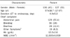
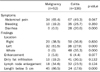
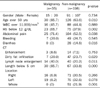
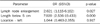
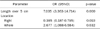
A total of 233 patients were included. The wall thickening was actually associated with abnormal endoscopic findings in 81.1% of the cases. The accuracy rate on diagnosis between CT and endoscopy was 63.5%. The discrepancy in diagnosis was higher in cases with left colon abnormality and short segment lesion. Abdominal pain was significantly more common in cases suspected malignancy on CT compared with colitis (p=0.047). Most of the malignancy diagnosed on CT involved the left side colon and most of the colitis involved the entire colon (p<0.001). The length of lesion was below 5 cm in 86.5% of the malignancy. Malignancy was more common in patients aged over 50 years with hemoglobin below 12 g/dL. The CT findings significantly suggestive of malignancy were lymph node enlargement and length of lesion below 5 cm (p=0.027 and p<0.001).
Conclusions
The large intestinal wall thickening on CT was limited in the differential diagnosis of malignancy and colitis. Additional endoscopic evaluation is needed in patients with bowel wall thickening associated with lymph node enlargement and short segment lesion on CT in order to exclude malignancy.
Figures and Tables
References
1. Nickoloff EL, Alderson PO. Radiation exposures to patients from CT: reality, public perception, and policy. AJR Am J Roentgenol. 2001. 177:285–287.
2. Prokop M. General principles of MDCT. Eur J Radiol. 2003. 45:Suppl 1. S4–S10.
3. Sebastian S, Kalra MK, Mittal P, Saini S, Small WC. Can independent coronal multiplanar reformatted images obtained using state-of-the-art MDCT scanners be used for primary interpretation of MDCT of the abdomen and pelvis? A feasibility study. Eur J Radiol. 2007. 64:439–446.
4. Coscina WF, Arger PH, Levine MS, et al. Gastrointestinal tract focal mass lesions: role of CT and barium evaluations. Radiology. 1986. 158:581–587.
5. Desai RK, Tagliabue JR, Wegryn SA, Einstein DM. CT evaluation of wall thickening in the alimentary tract. Radiographics. 1991. 11:771–783.
6. Griffiths JD. Surgical anatomy of the blood supply of the distal colon. Ann R Coll Surg Engl. 1956. 19:241–256.
7. Insko EK, Levine MS, Birnbaum BA, Jacobs JE. Benign and malignant lesions of the stomach: evaluation of CT criteria for differentiation. Radiology. 2003. 228:166–171.
8. Macari M, Balthazar EJ. CT of bowel wall thickening: significance and pitfalls of interpretation. AJR Am J Roentgenol. 2001. 176:1105–1116.
9. Wolff JH, Rubin A, Potter JD, et al. Clinical significance of colonoscopic findings associated with colonic thickening on computed tomography: is colonoscopy warranted when thickening is detected? J Clin Gastroenterol. 2008. 42:472–475.
10. Jeong JI, Park BC, Jeon WJ, et al. Clinical significance of bowel wall thickening detected with 64-slice multidetector computed tomography. Korean J Gastroenterol. 2009. 54:149–154.
11. Stermer E, Lavy A, Rainis T, Goldstein O, Keren D, Zeina AR. Incidental colorectal computed tomography abnormalities: would you send every patient for a colonoscopy? Can J Gastroenterol. 2008. 22:758–760.
12. Shin WC, Jeong MJ. Clinical significance of incidentally detected bowel wall thickening on abdominal computerized tomography scan. Korean J Gastroenterol. 2005. 45:409–416.
13. Eskaros S, Ghevariya V, Diamond I, Anand S. Correlation of incidental colorectal wall thickening at CT compared to colonoscopy. Emerg Radiol. 2009. 16:473–476.
14. Cai Q, Baumgarten DA, Affronti JP, Waring JP. Incidental findings of thickening luminal gastrointestinal organs on computed tomography: an absolute indication for endoscopy. Am J Gastroenterol. 2003. 98:1734–1737.
15. Balthazar EJ. CT of the gastrointestinal tract: principles and interpretation. AJR Am J Roentgenol. 1991. 156:23–32.
16. Tellez-Avila FI, García-Osogobio S, Chavez-Tapia NC, et al. Utility of endoscopy in patients with incidental gastrointestinal luminal wall thickening detected with CT. Surg Endosc. 2009. 23:2191–2196.
17. Low VH. The query corner. Bowel wall thickening on CT. Abdom Imaging. 1998. 23:107–110.
 XML Download