

 PDF
PDF ePub
ePub Citation
 Print
Print


Abstract
Background/Aims
Stomach cancer is prevalent in Korea. The purpose of this study was to evaluate the characteristics of superficial gastric cancers detected at SOK Sokpeynhan Internal Medical Network, the nationwide primary health care institutions.
Methods
We prospectively analysed the clinicopathologic and endoscopic characteristics of 218 superficial gastric cancer patients diagnosed using gastric endoscopy at SOK network from January 2011 through December 2011.
Results
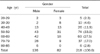
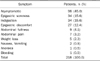
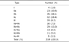
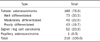
The mean age was 58.5 years old and male to female ratio was 1.7 : 1. Asymptomatic patients were most common (45.0%). The macroscopic classification revealed that simple types (63.8%) were more common than complex types (36.2%). The most common type was IIc (28.4%) and other types were as follows; IIb (16.1%), IIb+IIc (13.3%), IIa (10.6%), III (9.2%), IIa+IIc (7.3%), IIc+IIa (6.0%), IIc+IIb (5.0%). The most commonly involved sites were the body (53.1%) and greater curvature (32.6%) of the stomach. The size of lesion was less than 1 cm (69.3%) and less than 5 mm (33.5%) in diameter. The most common pathologic type was tubular adenocarcinoma (75.7%). Helicobacter pylori infection rate was 50.2%. Fifty five percent of the cases were diagnosed via endoscopy of National Health Insurance Corporation screenings.
Conclusions
Superficial gastric cancers in 2011 at primary health care SOK network were different from those of previous reports. Type IIc was most common but type IIb was more prevalent and the body and greater curvature of the stomach were the most commonly involved sites. Therefore, careful observation of the proximal gastric mucosa and mucosal color change is needed.
Figures and Tables

References
1. Korean Central Cancer Registry. The population based cancer incidence rate for 2009 in Korea. 2011. Ilsan: Korean Central Cancer Registry.
2. Lee HJ, Kim YH, Kim WH, et al. Clinicopathological analysis for recurrence of early gastric cancer. Jpn J Clin Oncol. 2003. 33:209–214.
3. The Paris endoscopic classification of superficial neoplastic lesions: esophagus, stomach, and colon: November 30 to December 1, 2002. Gastrointest Endosc. 2003. 58:6 Suppl. S3–S43.
4. Oota K, Sobin LH. Histological typing of gastric and oesophageal tumors. 1977. Geneva: WHO.
5. Sano T, Okuyama Y, Kobori O, Shimizu T, Morioka Y. Early gastric cancer. Endoscopic diagnosis of depth of invasion. Dig Dis Sci. 1990. 35:1340–1344.
6. Choi ES, Jeon WH, Sohn HJ, et al. Clinical study of early gastric carcinoma. Korean J Gastroenterol. 1995. 27:31–38.
7. Song HT, Kim CD, Ryu HS, Hyun JH. A clinical study of early gastric cancer. Korean J Gastroenterol. 1994. 26:789–799.
8. Kim TJ, Eom JW, Jeong JS, et al. Clinical study of early gastric cancer. Korean J Gastroenterol. 1993. 25:61–71.
9. Hisamichi S, Nozaki K, Kitagawa M, Mochizuki F, Gomi T. Evaluation of mass screening program for stomach cancer. Tohoku J Exp Med. 1976. 118:Suppl. 69–77.
10. Lee HJ, Chung JM, Seo EH, Jeon SW. Clinicopathologic characteristics of gastric cancer diagnosed at health screening. Korean J Med. 2008. 75:665–672.
11. Jeong HY, Lee SM, Lee KT, et al. Correspondence of endoscopic findings with histologic differentiation in early gastric cancer. Korean J Gastrointest Endosc. 2000. 20:83–90.
12. Park IS, Lee YC, Kim WH, Noh SH, Lee KS, Kim H. Clinicopathologic characteristics of early gastric cancer in Korea. Yonsei Med J. 2000. 41:607–614.
13. Kong SH, Park DJ, Lee HJ, et al. Clinicopathologic features of asymptomatic gastric adenocarcinoma patients in Korea. Jpn J Clin Oncol. 2004. 34:1–7.
14. Baek YH, Yoo HS, Yoon HA, et al. The usefulness of the endoscopic findings for predicting depth of invasion in early gastric cancer. Korean J Gastrointest Endosc. 2007. 35:297–303.
15. Kim JP, Hur YS, Yang HK. Lymph node metastasis as a significant prognostic factor in early gastric cancer: analysis of 1,136 early gastric cancers. Ann Surg Oncol. 1995. 2:308–313.
16. Tada M, Murakami A, Karita M, Yanai H, Okita K. Endoscopic resection of early gastric cancer. Endoscopy. 1993. 25:445–450.
17. Yoon SJ, Lee S, Lee OC, et al. A clinical study of early gastric cancer. Korean J Med. 1994. 47:381–387.
18. Kim HY, Cho BD, Chang WK, et al. Helicobacter pylori infection and the risk of gastric cancer among the Korean population. J Gastroenterol Hepatol. 1997. 12:100–103.
19. Chang WK, Kim HY, Kim YB, et al. Association between Helicobacter pylori infection and the risk of gastric cancer among the Korean people: prospective case-controlled study. Korean J Helicobacter Res Pract. 2002. 2:18–24.
20. Kim HY. What is the most important factor for gastric carcinogenesis in Koreans: Helicobacter pylori, host factor or environmental factor? Korean J Gastroenterol. 2007. 49:60–71.
21. Korean Central Cancer Registry. Cancer facts and figures 2011 in the Republic of Korea. 2011. Ilsan: Korean Central Cancer Registry.
 XML Download