

 PDF
PDF ePub
ePub Citation
 Print
Print


INTRODUCTION
Hepatitis A infection is caused by HAV, which is transmitted through the fecal-oral route. The incidence of adult hepatitis A has recently been increasing in Korea.1,2 The clinical manifestations of HAV infection depend on patient age: HAV infection is usually asymptomatic in children, while about 80% of infected adults experience severe hepatitis with remarkably elevated serum aminotransferases.3 Hepatitis A is most often a self-limited acute illness and does not progress to chronic hepatitis. In rare cases, however, acute hepatitis A is associated with serious complications such as fulminant hepatitis and acute kidney injury and may ultimately result in death or liver transplantation. In addition, hepatitis A is associated with various extrahepatic manifestations, such as hematologic abnormalities, pleural or pericardial effusion, acute reactive arthritis, acute pancreatitis, acalculous cholecystitis, mononeuritis, and Guillain-Barré syndrome.3
Pure red cell aplasia (PRCA) is a rare cause of anemia characterized by the absence of erythroid precursors in the bone marrow without changes in other cell lineages.4 Patients have severe anemia, low reticulocyte count, and normal platelet and granulocyte counts. PRCA is classified as acute or chronic and can be congenital or acquired. The acute form of PRCA is secondary to a virus- and drug-induced impairment of erythroid progenitor cells. The acquired chronic form of PRCA is associated with thymomas, lymphoproliferative disorders, autoimmune disorders, and immunocompromised states.4 PRCA is a rare hematopoietic complication of acute viral hepatitis, with very few reported cases of PRCA associated with HAV infection.5-9 Here, we report on our recent experience with a case of severe hepatitis A complicated by fulminant hepatitis and acute kidney injury followed by PRCA that a showed favorable response to oral corticosteroids.
CASE REPORT
A 39-year-old previously healthy woman was transferred to our hospital due to worsening jaundice, oliguria, and encephalopathy. She had been admitted to another hospital with general weakness and jaundice. The interval between the onset of jaundice and encephalopathy was 5 days. She had no known history of previous liver disease, heavy alcohol use, toxic agent exposure, or herbal medicine intake.
Vital signs were as follows: blood pressure 130/80 mmHg, pulse rate 86 beats/min, respiratory rate 20/min, body temperature 38.1℃. Physical examination showed generalized icterus, mild pretibial edema, and no hepatosplenomegaly. She had grade III hepatic encephalopathy with marked confusion and incoherent speech. She spent most of the time sleeping but could be aroused by vocal stimuli.
Laboratory tests on admission were as follows: hemoglobin (Hb) 11.6 g/dL, mean corpuscular volume 86.4, white blood cell (WBC) count 12.100/mm3, platelet count 98,000/mm3, erythrocyte sedimentation rate 25 mm/hour, CRP 23.5 mg/L, PT INR 1.56, AST 2,457 IU/L, ALT 4,176 IU/L, ALP 629 IU/L, GGT 430 IU/L, total bilirubin 4.1 mg/dL, direct bilirubin 3.2 mg/dL, total protein 6.1 g/dL, albumin 3.2 g/dL, ammonia 117 µmol/L (N: 11-32), cholesterol 97 mg/dL, glucose 136 mg/dL, BUN 93.5 mg/dL, creatinine 9.4 mg/dL, CK 841 IU/L (N: 32-187), LDH 1,111 IU/L (N: 218-472). Anti-HAV IgM and anti-HAV IgG were positive. Serological results for hepatitis B virus, hepatitis C virus, hepatitis E virus, human immunodeficiency virus, cytomegalovirus, and Epstein-Barr virus were negative. Serum copper and ceruloplasmin levels were normal. Anti-nuclear antibody, anti-dsDNA, anti-liver kidney microsome-1 antibody, and anti-smooth muscle antibody were negative. Urinalysis was notable for dark-colored urine with 1+ bilirubin, 3+ blood, and 1+ proteinuria. A chest roentgenogram initially showed mild pulmonary congestion, and abdominal ultrasonography was unremarkable.
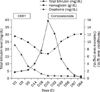
On the basis of the history and laboratory findings, acute viral hepatitis A complicated by acute kidney injury and fulminant hepatitis was diagnosed. She was admitted to the intensive care unit. Urine output was less than 30 mL/hour and did not increase with furosemide administration. With supportive care and close monitoring of hemodynamic and renal parameters, continuous renal replacement therapy was initiated. On the 5th hospital day, she was alert, and urine output had improved to within normal limits. Therefore, renal replacement therapy was stopped. On the 18th hospital day, despite improvements in serum AST, ALT, and creatinine levels, she showed a marked increase in bilirubin levels (total 19.4 mg/dL, direct bilirubin 14.4 mg/dL). In addition, Hb decreased to 6.9 g/dL with 0.14% reticulocytes; WBC and platelet counts were within normal limits. Peripheral blood smear revealed a normochromic, normocytic anemia. Other laboratory values were notable for serum ferritin >2,000 ng/mL, vitamin B12 1,134 pg/mL (N: 200-950), and folate 9.47 ng/mL (N: 3-17). Haptoglobin level was normal and a direct Coombs' test was negative. Parvovirus B19 IgM antibody was also negative. Stool occult blood was negative, and upper gastroendoscopy showed mild superficial gastritis. Bone marrow aspiration and biopsy showed a markedly elevated myeloid-erythroid ratio and the absence of red cell precursors with a normal range of myeloid and megakaryocytic cells (Fig. 1). The patient was diagnosed with viral hepatitis A-associated PRCA. She received repeated blood transfusions with no notable response. On the 25th hospital day, prednisone therapy was initiated at a dose of 1 mg/kg/day. Two weeks later, she gradually became transfusion-independent with a progressively increased Hb level (10.1 g/dL) and reticulocyte count (3.12%). In addition, total bilirubin level decreased, reaching 10.34 mg/dL. After 4 weeks of prednisolone therapy, it was tapered and stopped after 8 weeks of administration (Fig. 2). On the 24th weeks follow up, the patient remains asymptomatic with normal range of blood cell counts and liver function tests.
DISCUSSION
Improvements in the social and economic status and general public health of Korea over the last 30 years have changed the seroprevalance pattern of hepatitis A. Seropositivity for anti-HAV dropped to <10% in children and adolescents and 20-30% in young adults, while seropositivity is greater than 90% in adults over 40 years.10 These epidemiologic shifts led to a changing pattern of hepatitis A infection in Korea, ranging from an asymptomatic childhood infection to a increasing incidence of symptomatic disease in adults. Hepatitis A is most often a self-limited acute illness. However, it can rarely be complicated by fulminant hepatitis and acute kidney injury, resulting in death or liver transplantation. It has been suggested that factors influencing the severity of viral hepatitis A include chronic liver disease, increasing age, heavy alcohol consumption, human immunodeficiency virus infection, and pregnancy.11,12 It also has been very rarely associated with extrahepatic manifestations, such as hematologic abnormalities, pleural or pericardial effusion, acute reactive arthritis, acute pancreatitis, acalculous cholecystitis, mononeuritis, and Guillain-Barré syndrome.3 Many hematologic abnormalities can result from hepatitis A, including autoimmune hemolytic anemia, aplastic anemia, PRCA, and autoimmnue thrombocytopenia purpura. Hemolysis is precipitated by viral hepatitis, including hepatitis A, in patients with glucose-6-phosphate dehydrogenase deficiency.13 In addition, red cell survival in the absence of an underlying red cell abnormality can be shortended by acute infectious hepatitis (presumptive hepatitis A). Hemolysis may be autoimmune in nature, associated with antibodies to triphosphate isomerase, and can be severe. Other hematologic abnormalities include aplastic anemia, virus-associated hemophagocytic syndrome, autoimmune thrombocytopenic purpura, and PRCA.14,15
PRCA is a condition in which erythroid precursors in the bone marrow are nearly absent, while megakaryocytes and WBC precursors are present at normal levels.4 Erythroid precursors in the bone marrow are the primary targets in PRCA. As a result, patients can develop a normoblastic normochromic anemia and a virtual absence of reticulocytes. The acute form of PRCA is secondary to virus- and drug-induced impairment of erythroid progenitor cells. Respiratory infections, gastroenteritis, primary atypical pneumonia, infectious mononucleosis, mumps, and viral hepatitis may trigger PRCA. Most cases of acute transient PRCA are caused by parvovirus B19 infection. PRCA associated with acute hepatitis A is rare, with few cases having been reported.5-9 The mechanism of bone marrow suppression seen in many cased of viral hepatitis has not yet been elucidated. However, several possible mechanisms underlying the pathogenesis of PRCA associated with acute viral hepatitis have been suggested, including a direct effect of the hepatitis virus on erythropoiesis, the production of an antibody or inhibitor of erythropoietin, nutritional deficiency related to a hepatic disorder, and cell-mediated autoimmune suppression of erythropoiesis.7 Cytokines such as interleukin and circulating immune complexes are thought to be the triggers of erythropoiesis suppression.16
Initial management of PRCA involves the treatment of associated infections or other illness and discontinuation of offending drugs. An adequate Hb concentration (ranging from 8 to 10 g/dL) should be maintained with transfusion therapy. Treatment of PRCA usually includes corticosteroids or immunosuppressive or immunomodulating agents.17 Prednisone induces remission in approximately 45% of cases. In adults, prednisone therapy begins at 1-2 mg/kg per orally administered daily or 4-6 weeks and is tapered gradually when no longer indicated. If the underlying cause of PRCA is immunological and the response to corticosteroids has been inadequate, the next level of treatment is with cytotoxic or immunosuppressive drugs. Cyclophosphamide, 6-mercaptopurine, azathioprine, and cyclosporine have all been used. Plasmapheresis, autologous and nonmyeloablative allogeneic peripheral stem cell transplantation may be considered in patients who are refractory to other therapies. In the current case, following steroid administration, the level of Hb was increase to normal range within 8 weeks. There was no recurrence of hepatitis in our patient. During the administration of prednisolone, there were no complications or adverse events in association with the immunosuppression. It would be mandatory, however, to evaluate the possibility of recurrence of hepatitis following the discontinuation of prednisolone with monitoring of the clinical courses.
In conclusion, symptomatic hepatitis A in adults is currently increasing. Therefore, the incidence of more severe complications, such as fulminant hepatitis and acute kidney injury, is also expected to increase. Although rare, PRCA should be suspected in hepatitis A patients with anemia and low reticulocyte count along with a normal WBC and platelet count. Our patient, a 39-year-old woman, was diagnosed with acute hepatitis A complicated by fulminant hepatitis and acute kidney injury. In addition, she developed severe anemia with selective erythroid hypoplasia compatible with PRCA. Corticosteroid treatment was effective in improving clinical course of patient without replasing hepatitis.

 XML Download