

 PDF
PDF ePub
ePub Citation
 Print
Print


Abstract
Background/Aims
The usefulness of 18F-fluoro-2-deoxyglucose (FDG)-PET in detecting primary cancer, lymph node metastasis, and distant metastasis were studied in the gastric cancer patients.
Methods
The subjects were 392 gastric cancer patients who received FDG-PET and an abdominal CT test prior to surgery. The results of FDG-PET and CT were compared with the surgical and pathologic results.
Results
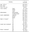
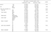
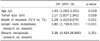
The primary site detection rate of FDG-PET was 74.4%, 50.3% in early gastric cancer and 92.0% in advanced gastric cancer. Detection rate was higher when tumors were larger than 3.5 cm, had deeper depth of invasion, and at a later stage (p<0.05, respectively). In multivariate analysis, tumor size, spread of tumor cells beyond the muscle layer (≥T2), and lymph node metastasis were statistically significant factors in primary site detection rate. The sensitivity, specificity, and positive predictive value of FDG-PET to lymph node metastasis were 59.6%, 88.8%, and 81.1% respectively, sensitivity being lower compared to CT while specificity and positive predictive value were higher. Sensitivity, specificity, and positive predictive value to distant metastasis were, respectively, 66.7%, 99.2%, and 88.0%, similar to CT. In 21 of the 392 patients (5.4%), synchronous double primary cancers were detected.
Figures and Tables
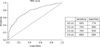
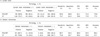
Fig. 1
Detection rate of gastric cancer with 18F-fluoro-2-deoxyglucose (FDG)-PET according to tumor size.
ROC, receiver operating characteristic.
Notes
References
1. Han EJ, Choi WH, Chung YA, et al. Comparison between FDG uptake and clinicopathologic and immunohistochemical parameters in pre-operative PET/CT scan of primary gastric carcinoma. Nucl Med Mol Imaging. 2009. 43:26–34.
2. Bar-Shalom R, Valdivia AY, Blaufox MD. PET imaging in oncology. Semin Nucl Med. 2000. 30:150–185.
3. Yeung HW, Macapinlac H, Karpeh M, Finn RD, Larson SM. Accuracy of FDG-PET in gastric cancer. Preliminary experience. Clin Positron Imaging. 1998. 1:213–221.
4. Koga H, Sasaki M, Kuwabara Y, et al. An analysis of the physiological FDG uptake pattern in the stomach. Ann Nucl Med. 2003. 17:733–738.
5. Terauchi T, Murano T, Daisaki H, et al. Evaluation of whole-body cancer screening using 18F-2-deoxy-2-fluoro-D-glucose positron emission tomography: a preliminary report. Ann Nucl Med. 2008. 22:379–385.
6. Kim SK, Kang KW, Lee JS, et al. Assessment of lymph node metastases using 18F-FDG PET in patients with advanced gastric cancer. Eur J Nucl Med Mol Imaging. 2006. 33:148–155.
7. Japanese Gastric Cancer Association. Japanese classification of gastric carcinoma-2nd English edition. Gastric Cancer. 1998. 1:10–24.
8. Hamilton SR, Aaltonen LA. Tumors of the stomach. WHO classification of tumors. Pathology and genetics. Tumors of the digestive system. 2000. Lyon: IARC Press;38–52.
9. Sobin LH, Wittekind C. International Union Against Cancer (UICC): TNM classification of malignant tumours. 2002. 6th ed. New York: Wiley-Liss;65–68.
10. Podoloff DA, Ball DW, Ben-Josef E, et al. NCCN task force: clinical utility of PET in a variety of tumor types. J Natl Compr Canc Netw. 2009. 7:Suppl 2. S1–S26.
11. Yamada A, Oguchi K, Fukushima M, Imai Y, Kadoya M. Evaluation of 2-deoxy-2-[18F]fluoro-D-glucose positron emission tomography in gastric carcinoma: relation to histological subtypes, depth of tumor invasion, and glucose transporter-1 expression. Ann Nucl Med. 2006. 20:597–604.
12. Stahl A, Ott K, Weber WA, et al. FDG PET imaging of locally advanced gastric carcinomas: correlation with endoscopic and histopathological findings. Eur J Nucl Med Mol Imaging. 2003. 30:288–295.
13. Chen J, Cheong JH, Yun MJ, et al. Improvement in preoperative staging of gastric adenocarcinoma with positron emission tomography. Cancer. 2005. 103:2383–2390.
14. Mukai K, Ishida Y, Okajima K, Isozaki H, Morimoto T, Nishiyama S. Usefulness of preoperative FDG-PET for detection of gastric cancer. Gastric Cancer. 2006. 9:192–196.
15. Mochiki E, Kuwano H, Katoh H, Asao T, Oriuchi N, Endo K. Evaluation of 18F-2-deoxy-2-fluoro-D-glucose positron emission tomography for gastric cancer. World J Surg. 2004. 28:247–253.
16. Peeters CF, de Waal RM, Wobbes T, Ruers TJ. Metastatic dormancy imposed by the primary tumor: does it exist in humans? Ann Surg Oncol. 2008. 15:3308–3315.
 XML Download