

 PDF
PDF ePub
ePub Citation
 Print
Print


Abstract
Background/Aims
We aimed to estimate the proportion of significant endoscopic findings and their association with dyspeptic symptoms and to evaluate the predictors for significant endoscopic findings.
Methods
Total of 3,872 subjects (58.3% men, mean age 43.6±9.3 years) who had undergone endoscopy were enrolled at the health promotion center. Each subject completed validated questionnaires, including data on gastrointestinal symptoms, socio-demographic history and medical history. Significant endoscopic findings were included peptic ulcer disease, reflux esophagitis, gastric cancer, Barrett's esophagus and gastro-duodenal erosions. Multiple logistic regression models were used to assess the predictors for significant endoscopic findings.
Results
The proportion of significant endoscopic findings was 39.1%. There was no significant difference of endoscopic findings between the dyspepsia and asymptomatic group (41.0% vs. 37.4%, p>0.05). There was no difference of the incidence of reflux esophagitis or peptic ulcer between subjects with and without dyspepsia. Peptic ulcer was more frequently present in subjects with reflux symptoms than asymptomatic subjects (12.3% vs. 9.0%, p=0.03). Male gender (odds ratio [OR], 3.91; 95% confidence interval [CI], 3.18-4.81) increased the risk for having endoscopic abnormality and having symptoms of functional dyspepsia according to Rome III criteria (OR, 0.75; 95% CI, 0.57-0.97) significantly decreased this risk.
Figures and Tables
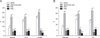 | Fig. 1Proportion of upper gastrointestinal (GI) symptoms according to reflux esophagitis, gastric ulcer and duodenal ulcer. (A) Upper GI symptoms in reflux esophagitis. (B) Upper GI symptoms in peptic ulcer disease.
GERS, gastroesophageal reflux symptoms, such as heartburn or acid regurgitation; EPS, epigastric pain syndrome; PDS, postprandial distress syndrome; LA-A, Los Angeles classification A; LA-B&C, Los Angeles classification B&C; GU, gastric ulcer, including active and healing stage ulcer; DU, duodenal ulcer, including active and healing stage ulcer.
aComparison of the proportion of GERD symptoms between GU vs. gastritis (8.9% vs. 14.9%, p=0.042).
|
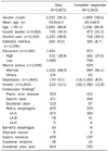
Table 2
Predictors for the Presence of Significant Endoscopic Findings by Multivariate Logistic Regression Analysis

![]()
Table 3
Significant Endoscopic Findings according to Upper Gastrointestinal Symptoms

Values are presented as n (%).
aDefined as having heartburn or acid regurgitation at least once a week; bdefined as having functional dyspepsia symptoms according to Rome III criteria; cincluded at least one as followings-active or healing stage of peptic ulcer disease, reflux esophagitis, Barrett esophagus, gastro-duodenal erosions, gastric cancer; d2 subjects had both gastric ulcer and duodenal ulcer; e2 subjects had both gastric ulcer and duodenal ulcer; freflux symptoms vs. no symptoms, p=0.03; gany dyspepsia symptoms vs. no symptoms, p=0.03; hreflux symptoms vs. no symptoms, p=0.04; endoscopic findings are not mutually exclusive.
![]()

References
1. Talley NJ, Silverstein MD, Agréus L, Nyrén O, Sonnenberg A, Holtmann G. American Gastroenterological Association. AGA technical review: evaluation of dyspepsia. Gastroenterology. 1998. 114:582–595.
2. Lam SK, Talley NJ. Report of the 1997 Asia Pacific Consensus Conference on the management of Helicobacter pylori infection. J Gastroenterol Hepatol. 1998. 13:1–12.
3. Kim JS, Lee KJ, Kim JH, Hahm KB, Cho SW. Functional gastrointestinal disorders in patients referred to specialist gastroenterologists in a tertiary hospital. Korean J Neurogastroenterol Motil. 2004. 10:111–117.
4. Talley NJ, Vakil NB, Moayyedi P. American gastroenterological association technical review on the evaluation of dyspepsia. Gastroenterology. 2005. 129:1756–1780.
5. Veldhuyzen van Zanten SJ, Bradette M, Chiba N, et al. Canadian Dyspepsia Working Group. Evidence-based recommendations for short- and long-term management of uninvestigated dyspepsia in primary care: an update of the Canadian Dyspepsia Working Group (CanDys) clinical management tool. Can J Gastroenterol. 2005. 19:285–303.
6. Kim JI, Kim SG, Kim N, et al. Korean College of Helicobacter and Upper Gastrointestinal Research. Changing prevalence of upper gastrointestinal disease in 28 893 Koreans from 1995 to 2005. Eur J Gastroenterol Hepatol. 2009. 21:787–793.
7. Jung HK, Na YJ, Moon IH. Changes of helicobacter pylori-positive peptic ulcer disease: based on data from a general hospital. Korean J Gastrointest Endosc. 2006. 32:1–8.
8. Han CH, Lee JS, Ahn JO, et al. The meaning of warning symptoms in the patients with dyspepsia. Korean J Med. 2007. 73:25–33.
9. Vakil N, Moayyedi P, Fennerty MB, Talley NJ. Limited value of alarm features in the diagnosis of upper gastrointestinal malignancy: systematic review and meta-analysis. Gastroenterology. 2006. 131:390–401.
10. Talley NJ, Phillips SF, Melton J 3rd, Wiltgen C, Zinsmeister AR. A patient questionnaire to identify bowel disease. Ann Intern Med. 1989. 111:671–674.
11. Talley NJ, Phillips SF, Wiltgen CM, Zinsmeister AR, Melton LJ 3rd. Assessment of functional gastrointestinal disease: the bowel disease questionnaire. Mayo Clin Proc. 1990. 65:1456–1479.
12. Song HJ, Jung HK, Yeom HJ, et al. Reliability and validity of Korean bowel disease questionnaire and prevalence of functional gastrointestinal disorders in Korea. Gut. 2009. 58:Suppl 1. A112.
13. Lundell LR, Dent J, Bennett JR, et al. Endoscopic assessment of oesophagitis: clinical and functional correlates and further validation of the Los Angeles classification. Gut. 1999. 45:172–180.
14. Sharma P, McQuaid K, Dent J, et al. AGA Chicago Workshop. A critical review of the diagnosis and management of Barrett's esophagus: the AGA Chicago Workshop. Gastroenterology. 2004. 127:310–330.
15. Correa P. Chronic gastritis: a clinico-pathological classification. Am J Gastroenterol. 1988. 83:504–509.
16. Dooley CP, Cohen H, Fitzgibbons PL, et al. Prevalence of Helicobacter pylori infection and histologic gastritis in asymptomatic persons. N Engl J Med. 1989. 321:1562–1566.
17. Tack J, Talley NJ, Camilleri M, et al. Functional gastroduodenal disorders. Gastroenterology. 2006. 130:1466–1479.
18. Barkun A, Leontiadis G. Systematic review of the symptom burden, quality of life impairment and costs associated with peptic ulcer disease. Am J Med. 2010. 123:358–366.
19. Zagari RM, Law GR, Fuccio L, Pozzato P, Forman D, Bazzoli F. Dyspeptic symptoms and endoscopic findings in the community: the Loiano-Monghidoro study. Am J Gastroenterol. 2010. 105:565–571.
 XML Download