

 PDF
PDF ePub
ePub Citation
 Print
Print


Case report
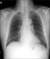
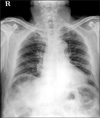
A 74-year-old man was admitted for endoscopic resection of laterally spreading tumor in the cecum, detected incidentally during health screening. His past medical history was remarkable for previous cholecystectomy due to calculous cholecystitis. On admission, physical examination and routine laboratory tests were unremarkable. Initial erect chest roentgenogram revealed no air under the right diaphragm (Fig. 1). The tumor was successfully resected by cap-assisted colonoscopic mucosal resection. He complained of abdominal discomfort and distention after colonoscopic mucosal resection. The physical examination revealed no guarding, tenderness or rebound tenderness. There was no tachypnea or tachycardia. Subsequent erect chest roentgenogram showed the right subdiaphragmatic air with haustral markings (Fig. 2). This finding was diagnosed as Chilaiditi's sign, characterized by interposition of the colon between liver and right diaphragm. Our case was managed conservatively and recovered.
Diagnosis: Chilaiditi's sign
Hepatodiaphragmatic interposition of the colon was first described by a Greek radiologist, Demetrius Chilaiditi1 in 1910. It is mostly diagnosed as an incidental finding on an erect chest or abdominal roentgenograms, which can be present temporarily or permanently. The incidence of this condition is reported as 0.025 to 0.28% in general population. It is more common in men and is much more in elderly and mentally retarded patients. This condition is generally asymptomatic, but may present with abdominal pain, nausea, vomiting, bloating, anorexia, diaphoresis, constipation, substernal pain, and even cardiac arrhythmias or respiratory distress.2-6 Less commonly, this condition has been associated with various abdominal complications including internal hernias, gastric and colonic volvulus, acute intestinal obstruction, and suprahepatic appendicitis.7,8 This is termed Chilaiditi's sign when asymptomatic and Chilaiditi's syndrome in symptomatic patients.
The predisposing factors of this condition remain unclear, but it may be predisposed in any factor that augments the space between the liver and diaphragm. These factors include the elongated, redundant and hypermobile colon (due to chronic constipation), laxity of suspensory ligaments, small liver (due to cirrhosis or hepatectomy), ascites, substantial weight loss in obese patients, abnormally high diaphragm (due to diaphragm muscular degeneration or phrenic nerve injury) and excessive aerophagia (in mental retarded patients).2-8
This condition can be confused with pneumoperitoneum and subphrenic abscess radiologically and can lead to unnecessary surgical intervention if not recognized correctly. Therefore, the understanding of the points of distinction between this condition and pneumoperitoneum and subphrenic abscess is important. The points of distinction are that haustral markings in subdiaphragmatic air are present and positional alteration of patient will not change the location of subdiaphragmatic air in hepatodiaphragmatic interposition of colon. However, if differential diagnosis is unclear, abdominal computed tomography is recommended to establish a definitive diagnosis.2,3,9-11
The management of this condition is often conservative approaches including bed rest, fluid supplementation, nasogastric decompression and laxatives, and surgical intervention has been occasionally indicated when symptoms worsened or when there was associated with various abdominal complications.11,12
In our case, the erect chest roentgenogram done after colonoscopic mucosal resection revealed the right subdiaphragmatic air with haustral markings. This finding was diagnosed as Chilaiditi's sign, characterized by hepatodiaphragmatic interposition of the colon. Our case was managed conservatively and recovered. The development of this condition in our case was thought to be due to excessive air insufflations during colonoscopic mucosal resection, which caused gaseous distention of the colon. As far as we are aware, this is the first report of Chilaiditi's sign induced by colonoscopic mucosal resection.
 XML Download