

 PDF
PDF ePub
ePub Citation
 Print
Print


INTRODUCTION
Hepatic metastases develop in about 50% of patients with colorectal cancer.1 Therefore, the management of patients with colorectal liver metastasis is a therapeutic challenge for surgeons and oncologists.
Therapeutic modalities can be divided into surgical resection, non-resectional ablation techniques, and regional or systemic chemotherapy. Of these, hepatic resection has been widely accepted as the optimal first-line modality for treating colorectal liver metastases.2,3 Hepatic resection is a potentially curative treatment for patients with hepatic metastases from colorectal carcinoma, and this carries a five-year survival rate of 32% to 58%.2,4-7 However, only about 10% of all patients with colorectal liver metastases are initial candidates for potentially curative resection.8
Among the non-resectional ablation techniques, radiofrequency ablation (RFA) is the most widely used modality for patients who are not candidates for hepatic resection.9 During the last decade, there has been considerable development in the RFA techniques for oncological applications.10 Therefore, the present study was performed to determine whether there were differences in outcome between RFA and surgical resection in the treatment of colorectal cancer with liver metastases.
SUBJECTS AND METHODS
Between July 2002 and April 2008, 25 patients underwent hepatic resection and 28 underwent RFA for synchronous or metachronous liver metastases of colorectal cancer at our hospital. Combination therapy of RFA and resection was excluded in the present study.
The diagnosis of liver metastasis was based on the findings from imaging studies such as ultrasonography and contrast material-enhanced CT with/without needle biopsy. Chest CT and/or PET-CT were performed to evaluate the extrahepatic disease. Needle aspiration biopsy was performed before treatment in only those patients recommended by the radiologist due to atypical hepatic mass enhancement. In the other patients, newly developed solid hepatic tumors with peripheral enhancement were considered to be hepatic metastases.
Our hospital had no predefined criteria of respectability with regard to either size or number of metastatic tumors. We considered hepatic resection whenever it was anticipated that the tumors could be completely resected (R0), more than two adjacent liver segments could be preserved with adequate vascular flow and biliary drainage, and the estimated liver volume following hepatic resection would be at least 20% of the total estimated liver volume. RFA was performed when co-morbidity, inadequate liver remnant or anatomical difficulty precluded hepatic resection or when patients refused the surgery due to risk of perioperative morbidity and mortality. Our inclusion criteria for performing RFA for colorectal liver metastases were as follows: (1) No signs of peritoneal dissemination or loco-regional disease or any other distant metastases, and (2) percutaneous complete ablations of the liver metastases were feasible. The study protocol was approved by the local ethics committee at our institute.
1. Hepatic resection and RFA techniques
Hepatic resection was performed simultaneously with colorectal surgery in 12 of 14 patients with synchronous liver metastasis. The other two patients with synchronous liver metastasis underwent delayed hepatic resection after neoadjuvant chemotherapy. Among 25 patients who underwent hepatic resection, 14 underwent anatomic hepatic resection of two and more segments, and additional non-anatomic resection was performed in five patients with a small tumor located in the other lobe. The other 11 patients underwent segmentectomy.
RFA of the hepatic metastases was performed percutaneously under local anesthesia by an interventional radiologist. Before RFA, all the patients were treated under intravenous conscious sedation. RFA was performed using a 200 W generator in the impedance control mode and a monopolar single or clustered internally cooled electrode with 3-cm exposure tip (Valleylab, Burlington, MA, USA). RFA was performed percutaneously under real-time sonographic guidance with a 3-5 MHz convex-array transducer (Prosound SSD-5500SV; Aloka, Tokyo, Japan). The radiofrequency current was applied for 12 minutes at 200 W to create a radius of ablation at least 10 mm larger than the largest tumor diameter. If the tumors were larger than 2.5 cm in the largest diameter, then we performed multiple overlapping ablations to cover the whole tumor plus a 5-mm ablative margin around the tumor. There was no formal written protocol in place for the type of electrode to use in ablation. In our experience, the advantage of a single electrode was ease of handling, while a clustered electrode enabled a larger volume of coagulation. A single electrode was adopted for tumors smaller than 3.0-3.5 cm, and a clustered electrode was used for larger or well-located tumors. After RFA, the electrode tract was cauterized to minimize bleeding.
2. After hepatic resection and RFA
Contrast enhanced CT studies were performed every three months after resection or RFA to evaluate for intrahepatic or extrahepatic recurrence. In patients who underwent RFA, the local therapeutic and technical efficacy were evaluated by additional contrast enhanced CT imaging one month after RFA, and the hypo-attenuating, non-enhancing areas observed during arterial and portal phases were considered to be indicative of complete tumor necrosis. Adjuvant chemotherapy was performed in 96.0% (24/25) of patients who underwent resection and 92.9% (26/28) of patients who underwent RFA. The patients in both groups suitable for intensive chemotherapy received the FOLFOX (5-FU, leucovorin and oxaliplatin) or FOLFIRI (5-FU, leucovorin and irinotecan) regimen. The patients in both groups not suited for intensive chemotherapy received oral chemoagents (doxifluridine or tegafur/uracil). The follow-up period ranged from 1.2 months to 62.0 months, with a median of 23.0 months.
3. Statistical analysis
All of the data are presented as median values with ranges. The significance of the differences between the results was tested using the chi-square test, Fisher's exact test, or Mann-Whitney test. A p-value<0.05 was considered statistically significant. Disease-free survival and overall survival after treatment were analyzed by the Kaplan-Meier method, and statistically significant differences in survival were identified by the log rank test. Factors associated with overall and disease-free survival were analyzed with multivariate analysis (type of treatment, detection time of metastases, number of tumors, size of main tumor) using the Cox proportional hazard model. Statistical analysis was performed with a statistical analysis program package (SPSS version 15.0; SPSS Inc., Chicago, IL, USA).
RESULTS
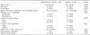
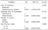
In the RFA and resection groups, the only oncologic clinical differences were CEA level and size of main tumors. The median CEA level at the time of diagnosis of liver metastases was significantly higher in the resection group (p=0.002), and the median size of the main liver metastases was significantly larger in the resection group (p=0.002) (Table 1).
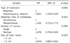
There was one major complication in each group, so there was no difference in the percentage of patients experiencing major complications. However, one patient who underwent right lobectomy for a 9.7-cm metastatic tumor died from multiple organ failure caused by postoperative hepatic failure. One patient who underwent RFA suffered from a 10-cm subcapsular hematoma, which was treated by conservative management for two months.
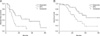
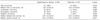
For outcomes after treatment, overall recurrence rate, median time to recurrence, intrahepatic recurrence, and extrahepatic recurrence, there were no differences between the two groups. However, the marginal recurrence rate was significantly higher in the RFA group (p=0.004) (Table 2). Disease-free survival was longer in the resection group (p=0.008), and overall survival was also longer in the resection group (p=0.017) (Fig. 1).
DISCUSSION
RFA has been used as a therapeutic alternative to hepatic resection for treating primary or metastatic tumors.11,12 During the last decade, RFA has been proposed as an alternative to hepatic resection for patients with limited hepatic involvement or solitary liver metastasis.13,14 However, recent studies have indicated that RFA was associated with a higher recurrence and a shorter disease-free survival.15,16 The present study also showed that the marginal recurrence rate was significantly higher in the RFA group, while disease-free and overall survival were significantly longer in the resection group. Local recurrence rates after RFA have been reported to range from 9% to 39%.7,17 In our previous study on RFA for hepatocellular carcinoma, although we tried to ablate the tumor as much as possible, small viable portions remained, and the percentage of viable portions tended to be higher as the tumor size increased.18 The resulting local recurrences could be reduced by surgical resection.
Recently, technical advances, increased experience and improvements in preoperative and postoperative care have led surgeons to expand the criteria for resectability in patients with colorectal liver metastases. The criteria for resectability have been expanded to include any patient with a tumor that can be removed with a negative margin and with adequate hepatic reserve.3 To date, although the majority of patients are not candidates for hepatic resection, it is a potentially curative treatment for hepatic metastases from colorectal carcinoma.2,3 Therefore, it may play a key role in improving the outcome of treatment to increase the resectability for colorectal liver metastases.
There are several methods for increasing resectability. First of all, neoadjuvant chemotherapies including oxaliplatin or/and irinotecan have led to higher resection rates (up to 38%) for unresectable colorectal liver metastases.19-22 Based on more recent data from a randomized phase II multicenter study (the CELIM study) of unresectable colorectal liver metastases that included targeted agents cetuximab, R0 resection rates after neoadjuvant chemotherapy were 38% (cetuximab plus FOLFOX6) and 30% (cetuximab plus FOLFIRI).23 Hepatic arterial infusion (HAI) chemotherapy also can be used to resect previously unresectable liver metastases. In one study, the resection rate was 47% after HAI with systemic chemotherapy for unresectable liver metastases.24
In addition to neoadjuvant chemotherapy, some surgical techniques to increase or preserve hepatic reserve volume have been introduced as a means to increase the resection rate. Portal vein embolization (PVE) or portal vein ligation (PVL) can be used to expand the resectability in patients who have a marginal estimated liver volume following hepatic resection.25 In patients with multiple and bilobar colorectal liver metastases, a two-staged hepatectomy with PVE or PVL may be the only potentially curative surgical approach with long-term survival.25,26 Combining hepatic resection with RFA also can expand the resectability as liver-directed surgical therapy; however survival was only slightly superior to that with nonsurgical treatment.7
The present study was retrospective in its data collection. There was possible bias in the selection of patients between the groups, because of variation in the severity of comorbidity, tumor size, and number of tumors. Another limitation of this study is the small number of cases. A study focusing on more optimal inclusion criteria of RFA and including a larger number of patients is needed to be performed in the future.
Until now, there have not been any randomized controlled trials comparing RFA to surgical resection for liver metastases from colorectal cancer, and most nonrandomized retrospective studies including the present study have shown inferior outcomes for RFA. Furthermore, there are many methods for increasing the resectability of colorectal liver metastases. Therefore, RFA for colorectal liver metastases should only be considered for selected patients with unresectable (by any means) disease or with high operative risk.
 XML Download