References
1. Hamano H, Kawa S, Horiuchi A, et al. High serum IgG4 concentrations in patients with sclerosing pancreatitis. N Engl J Med. 2001; 344:732–738.

2. Hamano H, Kawa S, Ochi Y, et al. Hydronephrosis associated with retroperitoneal fibrosis and sclerosing pancreatitis. Lancet. 2002; 359:1403–1404.
3. Kim KP, Kim MH, Song MH, Lee SS, Seo DW, Lee SK. Autoimmune chronic pancreatitis. Am J Gastroenterol. 2004; 99:1605–1616.

4. Yoshida K, Toki F, Takeuchi T, Watanabe S, Shiratori K, Hayashi N. Chronic pancreatitis caused by an autoimmune abnormality. Proposal of the concept of autoimmune pancreatitis. Dig Dis Sci. 1995; 40:1561–1568.
5. Zen Y, Harada K, Sasaki M, et al. IgG4-related sclerosing cholangitis with and without hepatic inflammatory pseudotumor, and sclerosing pancreatitis-associated sclerosing cholangitis: do they belong to a spectrum of sclerosing pancreatitis? Am J Surg Pathol. 2004; 28:1193–1203.
6. Taniguchi T, Ko M, Seko S, et al. Interstitial pneumonia associated with autoimmune pancreatitis. Gut. 2004; 53:770.
7. Tsushima K, Tanabe T, Yamamoto H, et al. Pulmonary involvement of autoimmune pancreatitis. Eur J Clin Invest. 2009; 39:714–722.

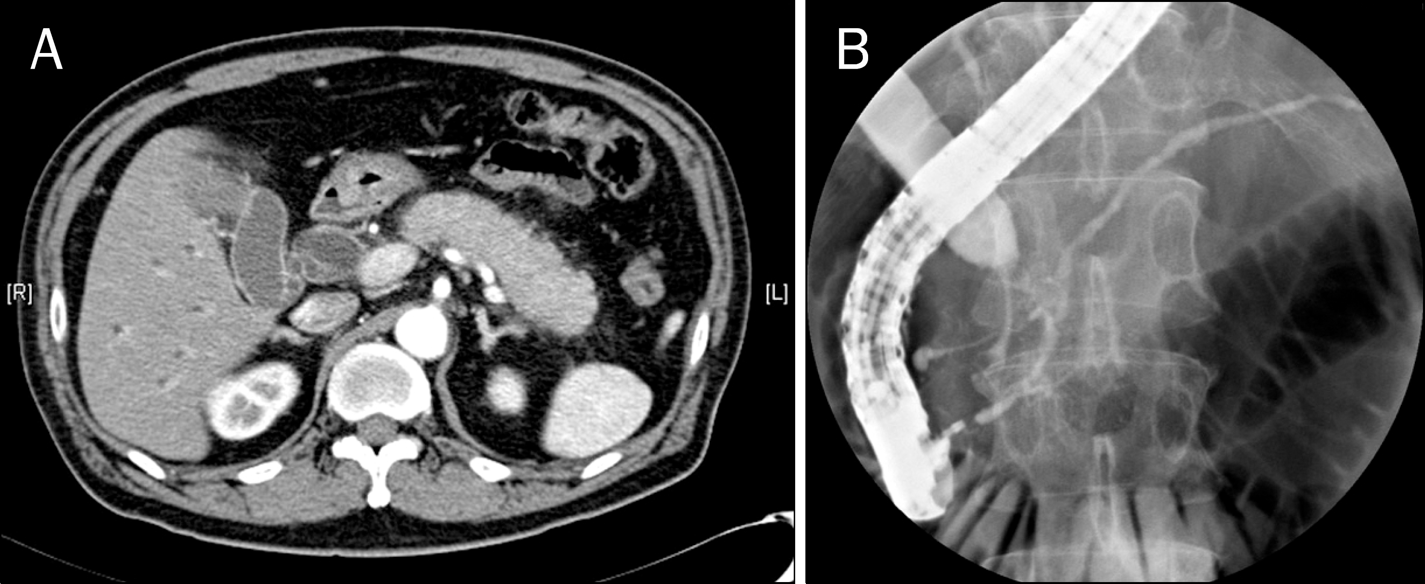
Fig. 1.
Abdominal CT and ERCP of autoimmune pancreatitis. (A) Abdominal CT showed diffuse enlargement of the pancreas, peripancreatic infiltration, and dilatation of the common bile duct, compatible with autoimmune pancreatitis. (B) ERCP showed obstruction of the distal common bile duct and diffuse slightly irregular pancreatic duct, compatible with autoimmune pancreatitis.

Fig. 2.
Chest CT of pulmonary involvement of autoimmune pancreatitis. (A) Chest CT showed multifocal consolidation with fibrosis and emphysematous change at both lungs. (B) Chest CT 1 month after azathioprine therapy showed the improvement of consolidation. (C) Chest CT 3 months after azathioprine therapy showed that previously noted consolidation disappeared completely.


 PDF
PDF ePub
ePub Citation
 Print
Print


 XML Download