References
1. Landry CS, Brock G, Scoggins CR, McMasters KM, Martin RC 2nd. A proposed staging system for rectal carcinoid tumors based on an analysis of 4701 patients. Surgery. 2008; 144:460–466.

2. Onaitis MW, Kirshbom PM, Hayward TZ, et al. Gastrointestinal carcinoids: characterization by site of origin and hormone production. Ann Surg. 2000; 232:549–556.

3. Modlin IM, Kidd M, Latich I, Zikusoka MN, Shapiro MD. Current status of gastrointestinal carcinoids. Gastroenterology. 2005; 128:1717–1751.

4. Sheikh H, Menakaya C, Hajdu L, Shackcloth M. Cystic degeneration of a carcinoid lung tumor: an uncommon cause of an intra-thoracic cyst. Ann Thorac Surg. 2011; 91:1966–1967.

5. Park YB, Kim JI, Ha BH, et al. Endoscopic treatment of gastrointestinal carcinoid tumors. Korean J Med. 2007; 73:274–282.
6. Sasaki Y, Niwa Y, Hirooka Y, et al. The use of endoscopic ultrasound-guided fine-needle aspiration for investigation of submucosal and extrinsic masses of the colon and rectum. Endoscopy. 2005; 37:154–160.

7. Loftus JP, van Heerden JA. Surgical management of gastrointestinal carcinoid tumors. Adv Surg. 1995; 28:317–336.
Fig. 1.
Colonoscopic finding. There was about 25 mm sized bulging mucosa with focal cystic portion in the rectum.
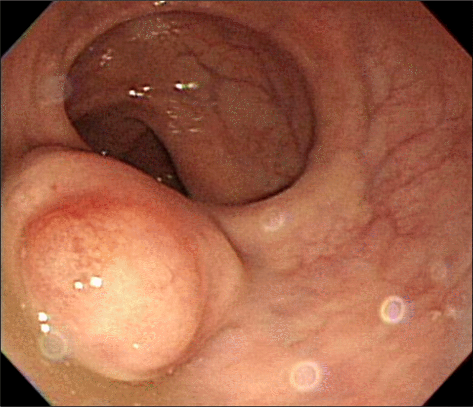
Fig. 2.
Endoscopic ultrasonography of the rectum. In the submucosal layer, about 2 cm sized hypoechoic mass was noted.
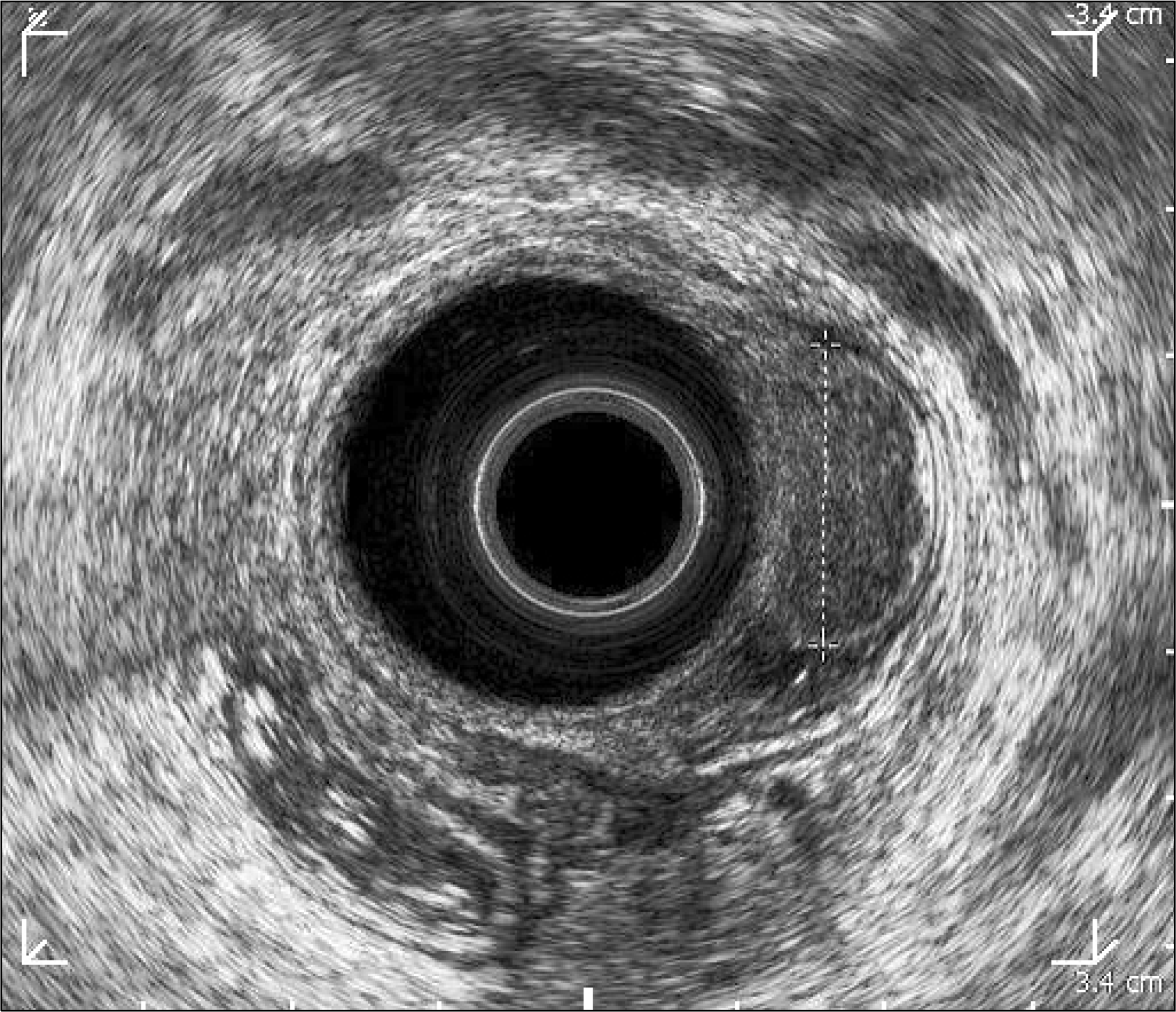
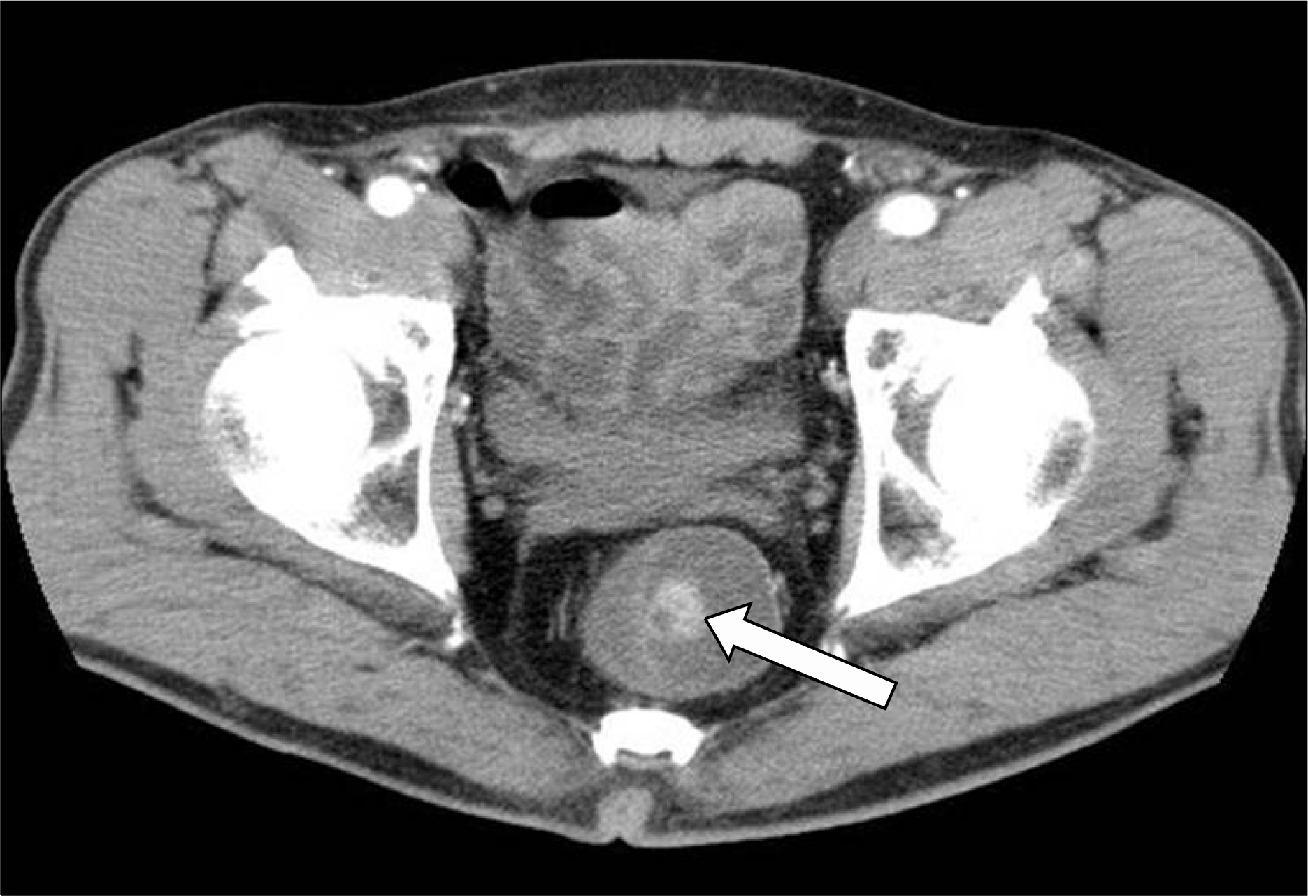
Fig. 3.
CT finding. About 2 cm sized polyphoid lesion (arrow) without adjacent organ infiltration was seen in the rectum.


 PDF
PDF ePub
ePub Citation
 Print
Print


 XML Download