

 PDF
PDF ePub
ePub Citation
 Print
Print


Abstract
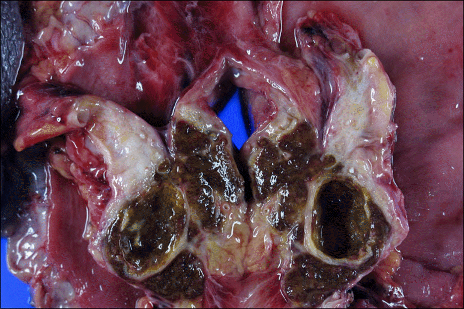
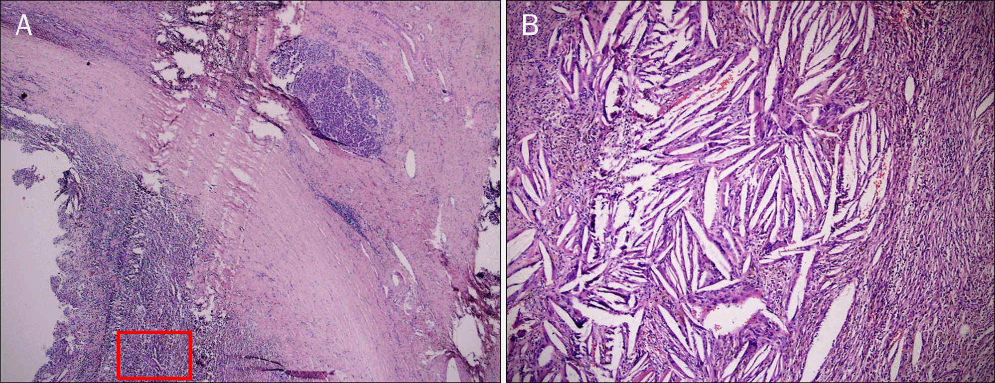
Cholesterol granuloma is a histological term used for the description of a tissue response to a foreign body such as cholesterol crystals. Cholesterol granuloma is histologically characterized as fibrous granulation tissue containing cholesterol crystals within surrounding giant cells. Cases of cholesterol granuloma of the pancreas are very rare. We report a case of a 47-year old male who had a cholesterol granuloma of the pancreas. Abdominal CT showed 24 mm-sized cyst in the pancreas and peripancreatic regional mass infiltrating to the stomach. PET-CT revealed increased 18F-FDG uptake at the cyst and peripancreatic mass. Thus, Whipple's operation was done. The disease was confirmed by surgical pathologic examination of the tissue. Pathologic examination of resected specimen showed numerous cholesterol crystals surrounded by multinucleated foreign body giant cells. We report on this case and give a brief review of the literature.
Go to : 
References
1. Ochiai H, Yamakawa Y, Fukushima T, Nakano S, Wakisaka S. Large cholesterol granuloma arising from the frontal si-nus–case report. Neurol Med Chir (Tokyo). 2001; 41:283–287.
2. Chin HS, Han DY, Jang WI, Lee IH, Kim YS, Lee BD. Clinical analysis of cholesterol granuloma in the middle ear. Korean J Otorhinolaryngol-Head Neck Surg. 2009; 52:974–979.

3. al-Amer AF, Walia HS, Madda JP. Cholesterol granuloma of the peritoneum. Can J Surg. 1990; 33:410–413.
4. Grignon DJ, Kirk ME, Haines DS. Cholesterol granulomas in lymph nodes draining a benign ovarian neoplasm. Arch Pathol Lab Med. 1985; 109:1124–1126.
5. Thevendran G, Al-Akraa M, Powis S, Davies N. Cholesterol granuloma of the kidney mimicking a tumour. Nephrol Dial Transplant. 2003; 18:2449–2450.
6. Reynolds HE, Cramer HM. Cholesterol granuloma of the breast: a mimic of carcinoma. Radiology. 1994; 191:249–250.
7. Heaton RB, Ross JJ, Jochum JM, Henry MR. Cytologic diagnosis of cholesterol granuloma. A case report. Acta Cytol. 1993; 37:713–716.
8. Klöppel G, Morohoshi T, John HD, et al. Solid and cystic acinar cell tumour of the pancreas. A tumour in young women with fa-vourable prognosis. Virchows Arch A Pathol Anat Histol. 1981; 392:171–183.
9. Manasse P. Ueber granulationsgeshwulst mit fremdkoerrie-senzellen. Virchows Arch. 1894; 136:245.
10. Chang JH, Lee SH, Chung WS. Three cases of orbitofrontal cholesterol granuloma. J Korean Ophthalmol Soc. 2005; 46:1228–1234.
11. Lee SW, Cha SH, Park DJ, Song GS, Choi CH, Lee YW. Cholesterol granuloma of frontal bone. J Korean Neurosurg Soc. 2001; 30:777–780.
12. Yoon WJ, Yoon YB, Lee KH, et al. The cystic neoplasms of the pancreas in Korea. Korean J Med. 2006; 70:261–267.
Go to :
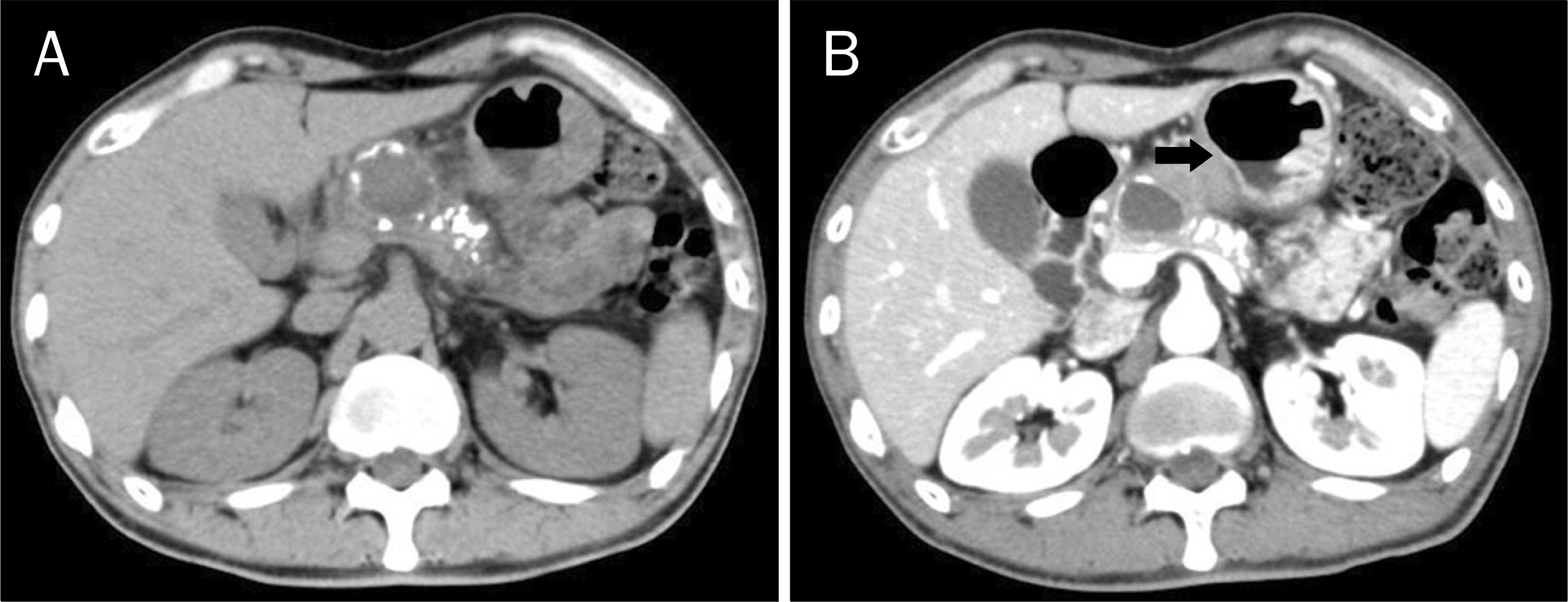 | Fig. 1.(A) Non-enhanced abdominal computerized tomography showed round cystic mass on the pancreas body portion with multiple peripheral rim calcifications. (B) Enhanced abdominal computerized tomography, 4 months later, showed diffuse thickening of the wall on the pancreas and the invasion of soft tissue mass to peripancreatic region (black arrow). |
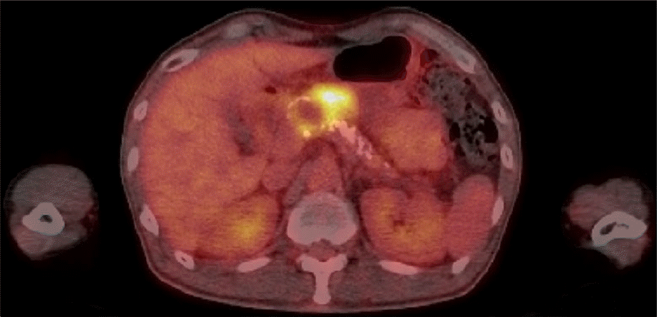 | Fig. 2.PET-CT showed increased 18F-FDG uptake in the rim of the cystic mass and peripancreatic mass. FDG, fluoro-deoxy-glucose. |
 XML Download