

 PDF
PDF ePub
ePub Citation
 Print
Print


Abstract
Duplication cysts are uncommon congenital malformations that may occur anywhere throughout the alimentary tract. The stomach is an extremely rare site of occurrence. Here, we report a case of gastric duplication cyst initially presenting with a gastric submucosal tumor. A 28-year-old man complained of dyspepsia lasting 1 year and upper endoscopy revealed an ellipsoid submucosal tumor at the greater curvature of the antrum. We intended to use the injection-and-cut technique: however, after saline injection, the lesion was dented and impossible to grasp with a snare. Therefore, we decided to perform endoscopic submucosal dissection and removed the tumor without complication. Histopathology revealed a 0.6×0.6 cm-sized duplication cyst, and there has been no recurrence in 2 years.
Go to : 
References
1. Kim DH, Kim JS, Nam ES, Shin HS. Foregut duplication cyst of the stomach. Pathol Int. 2000; 50:142–145.

2. Hsu HT, Hsing MT, Chen ML, Chen CJ. A gastric duplication cyst at the splenic hilum mimicking endometriosis clinically in a female adult. Chin Med J (Engl). 2009; 122:2079–2080.
3. Chen PH, Lee JY, Yang SF, Wang JY, Lin JY, Chang YT. A retroperitoneal gastric duplication cyst mimicking a simple exophytic renal cyst in an adolescent. J Pediatr Surg. 2010; 45:e5–e8.
4. Lee YC, Kim YB, Kim JK, et al. Endoscopic treatment of a large gastric duplication cyst with hook-knife and snare (with video). Gastrointest Endosc. 2011; 73:1039–1040.
5. Murakami S, Isozaki H, Shou T, Sakai K, Toyota H. Foregut duplication cyst of the stomach with pseudostratified columnar cili-ated epithelium. Pathol Int. 2008; 58:187–190.
6. Kuraoka K, Nakayama H, Kagawa T, Ichikawa T, Yasui W. Adeno-carcinoma arising from a gastric duplication cyst with invasion to the stomach: a case report with literature review. J Clin Pathol. 2004; 57:428–431.
7. Bonacci JL, Schlatter MG. Gastric duplication cyst: a unique presentation. J Pediatr Surg. 2008; 43:1203–1205.
8. Macpherson RI. Gastrointestinal tract duplications: clinical, pathologic, etiologic, and radiologic considerations. Radiographics. 1993; 13:1063–1080.
9. Parra-Blanco A, Arnau MR, Nicolás-Pérez D, et al. Endoscopic submucosal dissection training with pig models in a Western country. World J Gastroenterol. 2010; 16:2895–2900.
Go to :
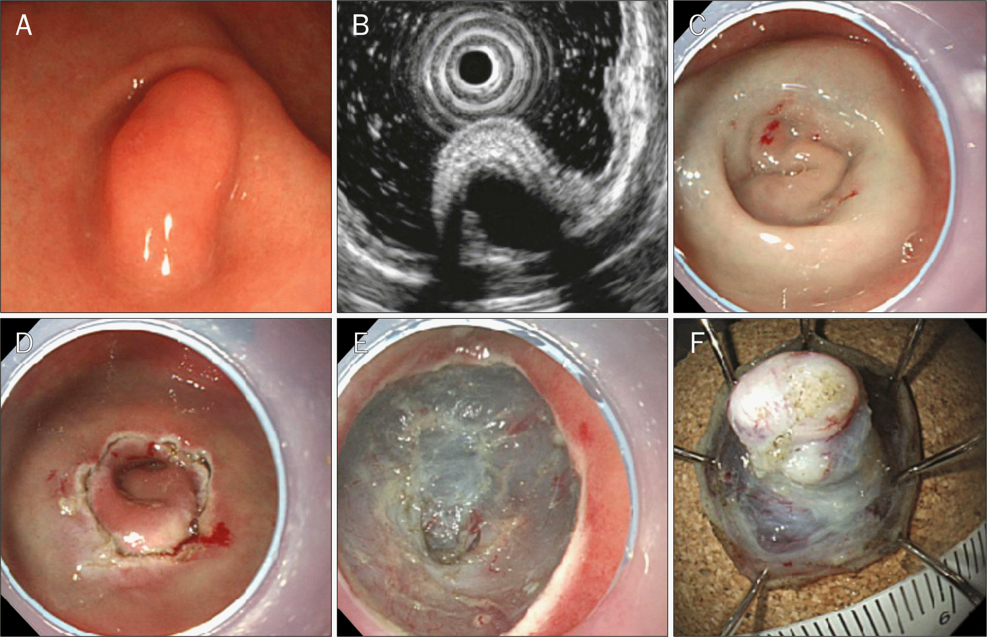 | Fig. 1.Endoscopic submucosal dissection of gastric duplication cyst. (A) A submucosal tumor at the greater curvature of the antrum. (B) EUS shows an anechoic homogenous, oval lesion originating from the submucosal layer of the stomach wall; the wall of the cystic lesion is shown as a five-layer structure. (C) After injecting saline with indigo carmine into the submucosa beneath the lesion, the lesion becomes flattened. (D) A complete circumference incision is made using an insulation-tipped knife. (E) The lesion is completely removed. (F) The inner surface of the resected specimen. |
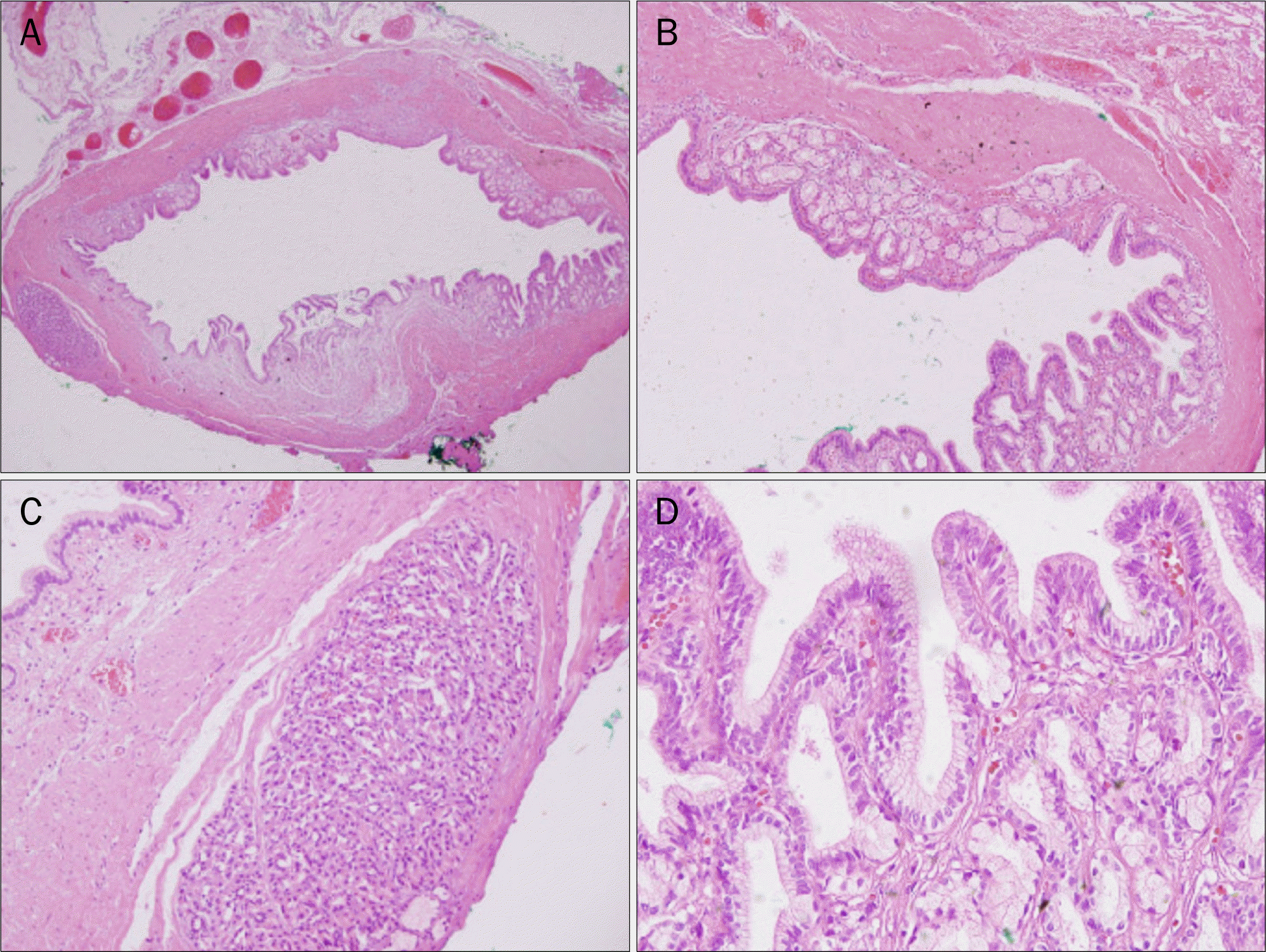 | Fig. 2.Histopathologic features of the resected specimen. (A) Pathologic examination revealed that the submucosal mass was a cystic lesion (H&E, ×20). (B) The cystic space was lined by columnar epithelial mucosa and had its own muscle layer (H&E, ×40). (C) In the cystic wall, ectopic pancreatic tissue was present, consisting of acinar and ductal structures (H&E, ×40). (D) The mucosa consisted of enteric- type columnnar epithelium (H&E, ×40). |
 XML Download