

 PDF
PDF ePub
ePub Citation
 Print
Print


Abstract
Background/Aims
Intraoperative cholangiogram (IOC) during laparoscopic cholecystectomy (LC) has been used to evaluate bile duct stone. But, the routine use of IOC remains controversial. With routine IOC during LC, we reviewed the variation of hepatic duct confluence and try to suggest the diagnostic criteria of asymptomatic common bile duct (CBD) stone.
Methods
We reviewed the medical record of 970 consecutive patients who underwent LC with IOC from January 1999 to December 2009, retrospectively.
Results
Nine hundered seventy patients were enrolled. IOC were successful in 957 (98.7%) and unsuccessful in 13 (1.3%). Eighty two of 957 patients (8.2%) were excluded because of no or poor radiologic image. According to Couinaud's classification, 492 patients (56.2%) had type A hepatic duct confluence, 227 patients (26.1%) type B, 15 patients (17%) type C1, 43 patients (4.9%) type C2, 72 patients (8.2%) type D1, 21 patients (2.4%) type D2, 1 patient (0.1%) type E1, 1 patient (0.1%) type E2, 2 patients (0.2%) type F, and 1 patient (0.1%) no classified type. The CBD stone was found in 116 of 970 (12.2%) patients. In 281 patients, preoperative serologic and radiologic tests did not show abnormality. When preoperative findings were not remarkable, there was no difference of clinical features between patients with or without CBD stones.
Go to : 
References
1. NIH Consensus Development Panel on gallstones and laparoscopic cholecystectomy. Gallstones and laparoscopic cholecystectomy. Surg Endosc. 1993; 7:271–279.
2. Jones DB, Dunnegan DL, Soper NJ. Results of a change to routine fluorocholangiography during laparoscopic cholecystectomy. Surgery. 1995; 118:693–701.

3. Woods MS, Traverso LW, Kozarek RA, et al. Biliary tract complications of laparoscopic cholecystectomy are detected more frequently with routine intraoperative cholangiography. Surg Endosc. 1995; 9:1076–1080.
4. Soper NJ, Dunnegan DL. Routine versus selective intraoperative cholangiography during laparoscopic cholecystectomy. World J Surg. 1992; 16:1133–1140.
5. Hauer-Jensen M, Karesen R, Nygaard K, et al. Prospective randomized study of routine intraoperative cholangiography during open cholecystectomy: long-term follow-up and multivariate analysis of predictors of choledocholithiasis. Surgery. 1993; 113:318–323.
6. Kullman E, Borch K, Lindström E, Svanvik J, Anderberg B. Value of routine intraoperative cholangiography in detecting aberrant bile ducts and bile duct injuries during laparoscopic cholecystectomy. Br J Surg. 1996; 83:171–175.
7. Stuart SA, Simpson TI, Alvord LA, Williams MD. Routine intraoperative laparoscopic cholangiography. Am J Surg. 1998; 176:632–637.
8. Snow LL, Weinstein LS, Hannon JK, Lane DR. Evaluation of operative cholangiography in 2043 patients undergoing laparoscopic cholecystectomy: a case for the selective operative cholangiogram. Surg Endosc. 2001; 15:14–20.
9. Couinaud C, Le FOI. Études anatomiques et chirurgicales. Paris: Masson;1957.
10. Kune GA. The influence of structure and function in the surgery of the biliary tract. Ann R Coll Surg Engl. 1970; 47:78–91.
11. Wang HP, Wu MS, Lin CC, et al. Pancreaticobiliary diseases associated with anomalous pancreaticobiliary ductal union. Gastrointest Endosc. 1998; 48:184–189.
12. Mirizzi PL. La cholangiografia durante las operaciones de las vias biliares. Bol Soc Cir Buenos Aires. 1932; 16:1133.
13. Hicken NF, Best RR, Hunt HB. Cholangiography: Visualization of the gallbladder and bile ducts during and after operation. Ann Surg. 1936; 103:210–229.
14. Kakos GS, Tompkins RK, Turnipseed W, Zollinger RM. Operative cholangiography during routine cholecystectomy: a review of 3,012 cases. Arch Surg. 1972; 104:484–488.
15. Berci G, Hamlin JA. Operative biliary radiology. Baltimore: Williams & Wilkins;1981.
16. McSherry CK. Cholecystectomy: the golden standard. Am J Surg. 1989; 158:174–178.
17. Kuster GG, Gilroy S, Graefen M. Intraoperative cholangiography for laparoscopic cholecystectomy. Surg Gynecol Obstet. 1993; 176:411–417.
18. Choi JW, Kim TK, Kim KW, et al. Anatomic variation in intrahepatic bile ducts: an analysis of intraoperative cholangio-grams in 300 consecutive donors for living donor liver transplantation. Korean J Radiol. 2003; 4:85–90.
19. Flowers JL, Zucker KA, Graham SM, Scovill WA, Imbembo AL, Bailey RW. Laparoscopic cholangiography. Results and indications. Ann Surg. 1992; 215:209–216.
20. Rhodes M, Sussman L, Cohen L, Lewis MP. Randomised trial of laparoscopic exploration of common bile duct versus post-operative endoscopic retrograde cholangiography for common bile duct stones. Lancet. 1998; 351:159–161.
21. Erickson RA, Carlson B. The role of endoscopic retrograde cholangiopancreatography in patients with laparoscopic chole-cystectomies. Gastroenterology. 1995; 109:252–263.
22. Phillips EH, Berci G, Carroll B, Daykhovsky L, Sackier J, Paz- Partlow M. The importance of intraoperative cholangiography during laparoscopic cholecystectomy. Am Surg. 1990; 56:792–795.
23. Ammori BJ, Birbas K, Davides D, Vezakis A, Larvin M, McMahon MJ. Routine vs "on demand" postoperative ERCP for small bile duct calculi detected at intraoperative cholangiography. Clinical evaluation and cost analysis. Surg Endosc. 2000; 14:1123–1126.
24. Nickkholgh A, Soltaniyekta S, Kalbasi H. Routine versus selective intraoperative cholangiography during laparoscopic cholecystectomy: a survey of 2,130 patients undergoing laparoscopic cholecystectomy. Surg Endosc. 2006; 20:868–874.
25. Mills JL, Beck DE, Harford FJ Jr. Routine operative cholangiography. Surg Gynecol Obstet. 1985; 161:343–345.
26. Kitahama A, Kerstein MD, Overby JL, Kappelman MD, Webb WR. Routine intraoperative cholangiogram. Surg Gynecol Obstet. 1986; 162:317–322.
Go to :
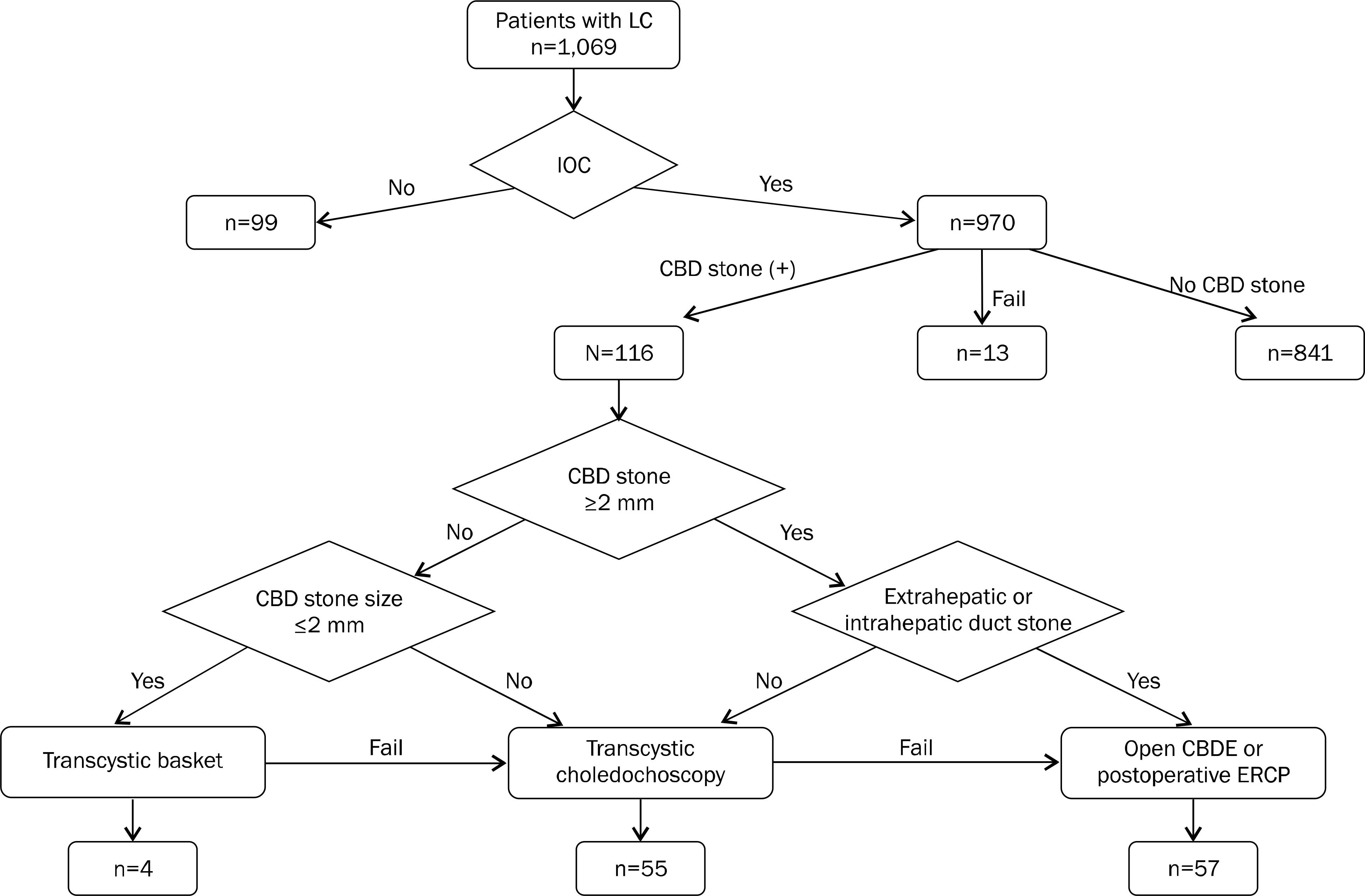 | Fig. 1.IOC flow chart. It shows the process of IOC and how to confirm CBD stone defected with IOC. IOC, intraoperative cholangiogram; CBD, common bile duct; LC, laparoscopic cholecystectomy. |
Table 1.
Reasons for Not Doing Intraoperative Cholangiogram
Table 2.
Reasons for Conversion to Open Cholecystectomy
| N (%) | |
|---|---|
| Adhesion | 41 (86) |
| Failed cystic duct ligation | 2 (4) |
| Cystic duct injury | 1 (2) |
| Failed choledochoscopic removal of CBD stone | 1 (2) |
| CBD injury | 2 (4) |
| Malignancy | 1 (2) |
| Total | 48 (100) |
Table 3.
Cross Data between IHD Type and Cystic Duct Confluence Position
Table 4.
Anatomincal Variation in Intraoperative Cholangiogram
| n (%) | |
|---|---|
| Pancreatic opacification | 117 |
| Anomaly of pancreaticobiliary duct union | 12 |
| Choledochal cyst | 2 |
| Others | 6 |
| Total | 137 |
Table 5.
Results of Intraoperative Cholangiogram
| n (%) | |
|---|---|
| True positive | 96 (9.9) |
| True negative | 852 (87.8) |
| False positive | 20 (2.1) |
| False negative | 2 (0.2) |
| Total | 970 (100) |
Table 6.
Comparative Analysis of Asymptomatic Common Bile Duct Stone
| Group 1 (n=12) | Group 2 (n=269) | p-value | |
|---|---|---|---|
| Age (mean, yr) | 54.6 | 50.62 | .478 a |
| BMI (mean) | 24 | 24.5 | .670 a |
| ASA (I: II: III, %) | 58:25:17 | 77:21:2 | .730 b |
| Gender (male: female, %) | 42:58 | 40:60 | .417 b |
| Period from test to IOC (mean, day) | 1.75 | 1.98 | .912 a |
| GB stone number (mean) | 5.38 | 4.41 | .467 a |
| GB stone size (mean, mm) | 10.13 | 9.07 | .752 a |
| GB wall thickness (mean, mm) | 4.0 | 3.56 | .568 a |
 XML Download