

 PDF
PDF ePub
ePub Citation
 Print
Print


References
1. Anthony PP. Primary carcinoma of the liver: a study of 282 cases in Ugandan Africans. J Pathol. 1973; 110:37–48.

2. Nozaki Y, Kobayashi N, Shimamura T, et al. Colonic metastasis from hepatocellular carcinoma: manifested by gastrointestinal bleeding. Dig Dis Sci. 2008; 53:3265–3266.
3. Lin CP, Cheng JS, Lai KH, et al. Gastrointestinal metastasis in hepatocellular carcinoma: radiological and endoscopic studies of 11 cases. J Gastroenterol Hepatol. 2000; 15:536–541.
4. Chen LT, Chen CY, Jan CM, et al. Gastrointestinal tract involvement in hepatocellular carcinoma: clinical, radiological and endoscopic studies. Endoscopy. 1990; 22:118–123.
5. Nakashima T, Okuda K, Kojiro M, et al. Pathology of hepatocellular carcinoma in Japan. 232 Consecutive cases autopsied in ten years. Cancer. 1983; 51:863–877.
6. Park MS, Kim KW, Yu JS, et al. Radiologic findings of gastrointestinal tract involvement in hepatocellular carcinoma. J Comput Assist Tomogr. 2002; 26:95–101.
7. Yoo DJ, Chung YH, Lee YS, et al. Sigmoid colon metastasis from hepatocellular carcinoma. Korean J Hepatol. 2010; 16:397–400.
8. Yang PM, Sheu JC, Yang TH, et al. Metastasis of hepatocellular carcinoma to the proximal jejunum manifested by occult gastrointestinal bleeding. Am J Gastroenterol. 1987; 82:165–167.
9. Goo JC, Kim BU, Jeong JI, et al. Clinical observation after resection of lower gastrointestinal carcinoid tumor. Intest Res. 2010; 8:142–150.
10. Choi SH, Kim SJ, Choi YJ, et al. Clinicopathologic analysis of gastrointestinal stromal tumors of the colon and rectum. J Korean Soc Coloproctol. 2009; 25:323–333.
11. Yang SK, Byeon JS. Colonoscopy: diagnosis and treatment. 2nd ed.Seoul: Koonja;2009.
12. Katon RM, Brendler SJ, Ireland K. Gastric linitis plastica with metastases to the colon: a mimic of Crohn's disease. J Clin Gastroenterol. 1989; 11:555–560.
13. Koelma IA, Nap M, Huitema S, Krom RA, Houthoff HJ. Hepatocellular carcinoma, adenoma, and focal nodular hyperplasia. Comparative histopathologic study with immunohistochemical parameters. Arch Pathol Lab Med. 1986; 110:1035–1040.
14. Kojiro M, Kawano Y, Isomura T, Nakashima T. Distribution of al-bumin- and/or alpha-fetoprotein-positive cells in hepatocellular carcinoma. Lab Invest. 1981; 44:221–226.
15. Kakizoe S, Kojiro M, Nakashima T. Hepatocellular carcinoma with sarcomatous change. Clinicopathologic and immunohistochemical studies of 14 autopsy cases. Cancer. 1987; 59:310–316.
16. Seok JY, Kim YB. Sarcomatoid hepatocellular carcinoma. Korean J Hepatol. 2010; 16:89–94.
17. Maeda T, Adachi E, Kajiyama K, Takenaka K, Sugimachi K, Tsuneyoshi M. Spindle cell hepatocellular carcinoma. A clinicopathologic and immunohistochemical analysis of 15 cases. Cancer. 1996; 77:51–57.
18. Roskams T, Kojiro M. Pathology of Hepatocellular Carcinoma. Oxford: Blackwell Publishing;2006. p. 88–89.
19. Natsuizaka M, Omura T, Akaike T, et al. Clinical features of hepatocellular carcinoma with extrahepatic metastases. J Gastroenterol Hepatol. 2005; 20:1781–1787.
Go to : 
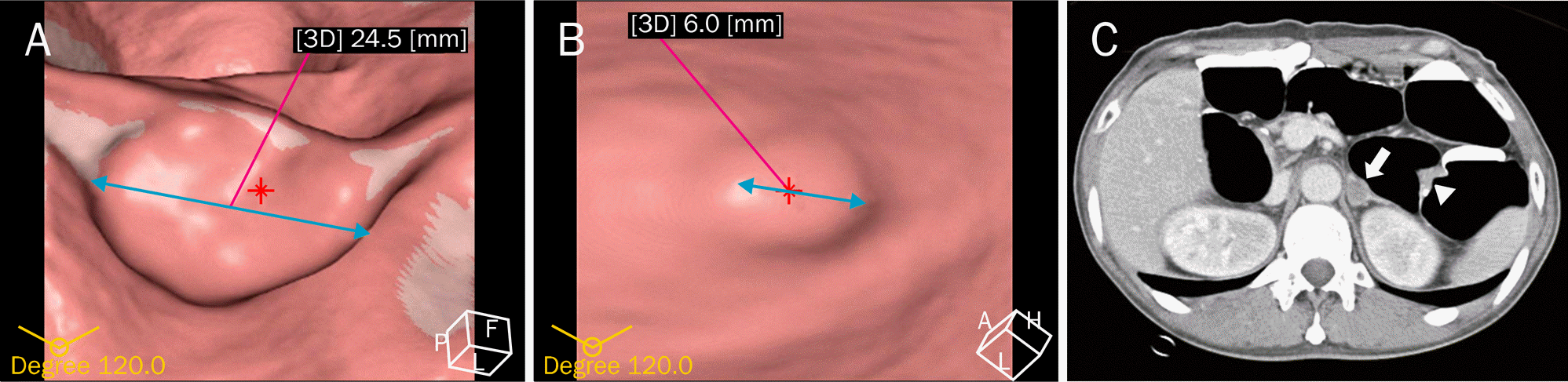 | Fig. 1.CT virtual colonoscopy. (A) A 24.5 mm-sized ulcerofungating mass was observed in the proximal descending colon. (B) A 6 mm-sized sessile polyp was also noted in the descending colon. (C) A 15 mm-sized low attenuated nodule suggesting metastatic tumor was noted in the left adrenal gland (arrow). Ulcerofungating mass in the proximal descending colon was also noted (arrowhead). |
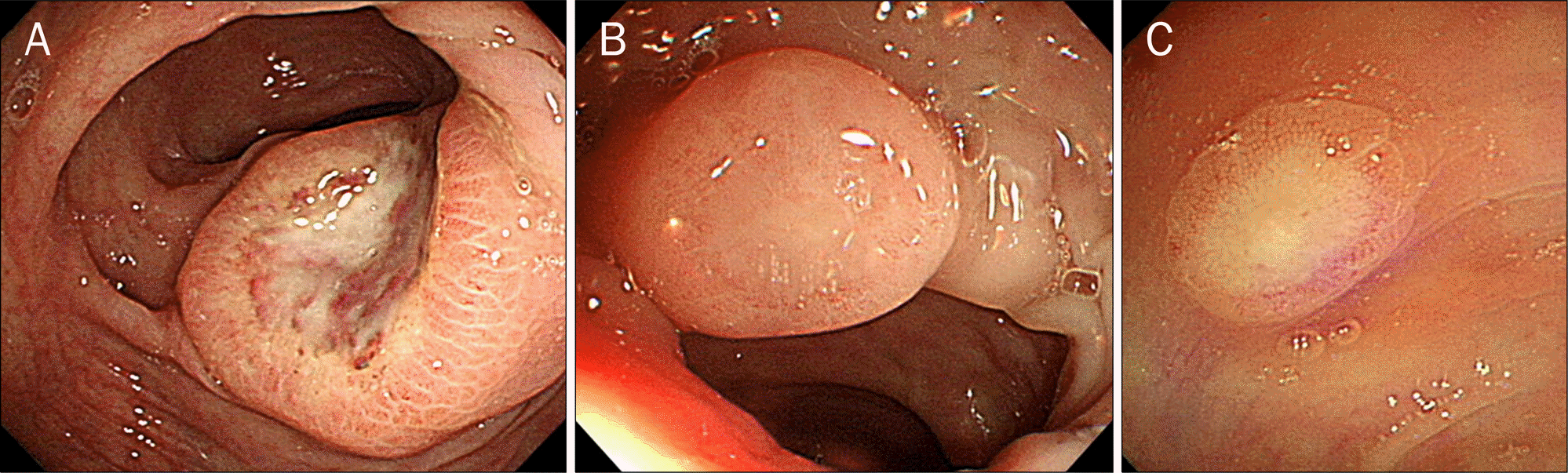 | Fig. 2.Colonoscopic findings. (A) An about 25 mm-sized ovoid mass with central ulceration was noted in the proximal descending colon. The mucosa around ulceration showed similar feature compared with normal surrounding colonic mucosa. (B, C) Smaller tumors with similar morphologies with (A) were noted in the hepatic flexure and the descending colon. |
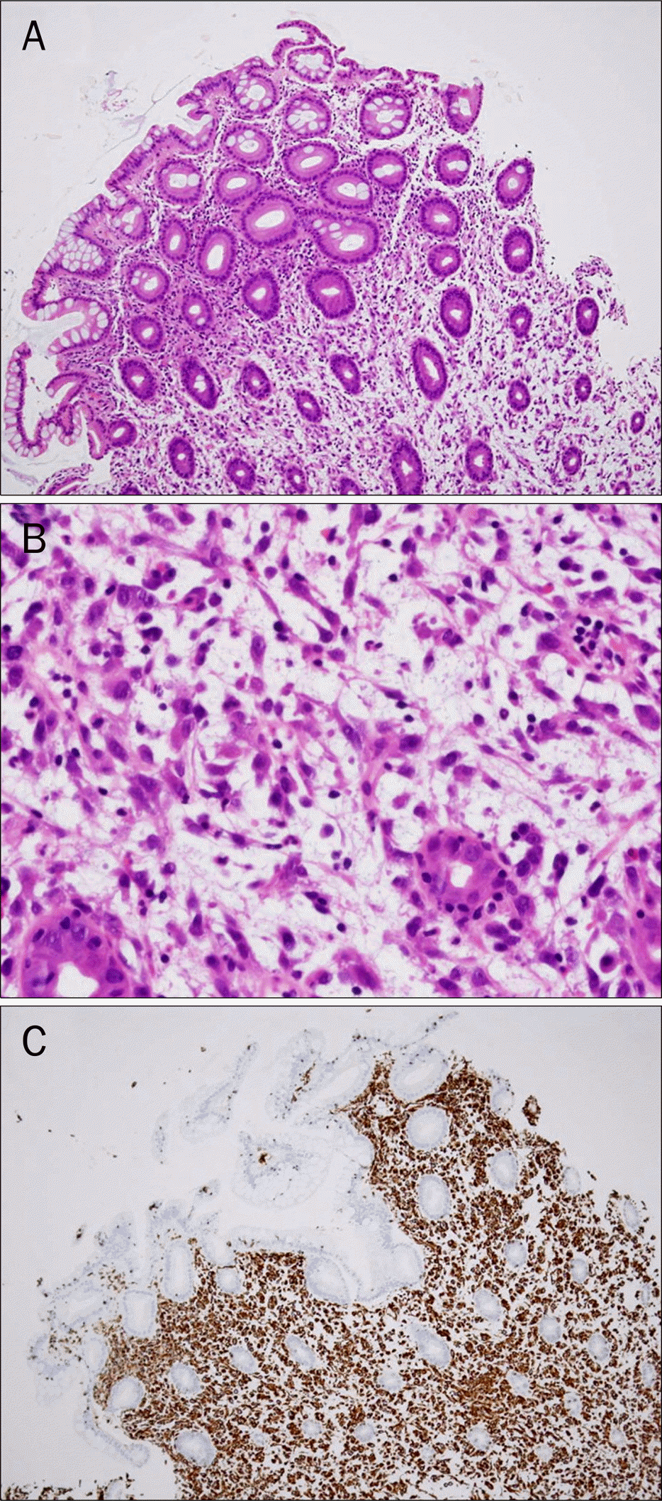 | Fig. 3.Histologic findings of metastatic sarcoid hepatocellular carcinoma. (A) The colonic crypts were normal and tumor cells infiltrating lamina propria were noted (H&E, ×100). (B) The tumor cells which had large, irregular and pale nuclei and spindle shaped cytoplasm were loosely infiltrating the lamina propria (H&E, ×400).(C) The tumor cells infiltrating the lamina propria were positive for vimentin (×100). |

 XML Download