

 PDF
PDF ePub
ePub Citation
 Print
Print


Abstract
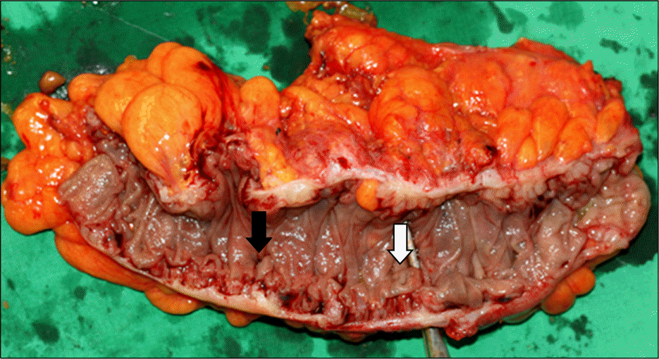
Enterovesical fistular is an abnormal communication between the intestine and the bladder. It represents a rare complication of intestinal diverticulitis, colorectal malignancy, bladder cancer, inflammatory bowel disease, radiotherapy, and trauma. The most common etiology is diverticular disease. A 70-year-old man came to our hospital due to frequent urinary tract infection, dysuria, pneumaturia and fecaluria. Sigmoidoscopy revealed a large diverticulum with impacted stool at the sigmoid colon. When the scope was inserted into the site, the patient complained of severe urgency and pneumaturia. CT scan was performed. 1.5 cm sized fistular tract between the sigmoid colon and bladder was noted. According to the endoscopy and CT finding, the diagnosis of colovesical fistula was made. The patient underwent surgical intervention. At laparotomy, there were multiple diverticula and fistular tract was noted.
Go to : 
References
1. Young-Fadok TM, Roberts PL, Spencer MP, Wolff BG. Colonic diverticular disease. Curr Probl Surg. 2000; 37:457–514.

2. Kavanagh D, Neary P, Dodd JD, Sheahan KM, O'Donoghue D, Hyland JM. Diagnosis and treatment of enterovesical fistulae. Colorectal Dis. 2005; 7:286–291.
3. Daniels IR, Bekdash B, Scott HJ, Marks CG, Donaldson DR. Diagnostic lessons learnt from a series of enterovesical fistulae. Colorectal Dis. 2002; 4:459–462.
4. Pollard SG, Macfarlane R, Greatorex R, Everett WG, Hartfall WG. Colovesical fistula. Ann R Coll Surg Engl. 1987; 69:163–165.
5. Najjar SF, Jamal MK, Savas JF, Miller TA. The spectrum of colovesical fistula and diagnostic paradigm. Am J Surg. 2004; 188:617–621.
6. Solkar MH, Forshaw MJ, Sankararajah D, Stewart M, Parker MC. Colovesical fistula-is a surgical approach always justified? Colorectal Dis. 2005; 7:467–471.
7. Shatila AH, Ackerman NB. Diagnosis and management of colovesical fistulas. Surg Gynecol Obstet. 1976; 143:71–74.
8. Bahadursingh AM, Virgo KS, Kaminski DL, Longo WE. Spectrum of disease and outcome of complicated diverticular disease. Am J Surg. 2003; 186:696–701.
9. Cripps H. Gow: passage of gas and faeces through the urethra: colostomy, recovery, remarks. Lancet. 1888; 2:619–620.
10. Garcea G, Majid I, Sutton CD, Pattenden CJ, Thomas WM. Diagnosis and management of colovesical fistulae; six-year experience of 90 consecutive cases. Colorectal Dis. 2006; 8:347–352.
11. Melchior S, Cudovic D, Jones J, Thomas C, Gillitzer R, Thüroff J. Diagnosis and surgical management of colovesical fistulas due to sigmoid diverticulitis. J Urol. 2009; 182:978–982.
12. Ferguson GG, Lee EW, Hunt SR, Ridley CH, Brandes SB. Management of the bladder during surgical treatment of enterovesical fistulas from benign bowel disease. J Am Coll Surg. 2008; 207:569–572.
13. Hsieh JH, Chen WS, Jiang JK, Lin TC, Lin JK, Hsu H. Enterovesical fistula: 10 years experience. Zhonghua Yi Xue Za Zhi (Taipei). 1997; 59:283–288.
14. Kwon EO, Armenakas NA, Scharf SC, Panagopoulos G, Fracchia JA. The poppy seed test for colovesical fistula: big bang, little bucks! J Urol. 2008; 179:1425–1427.
Go to :
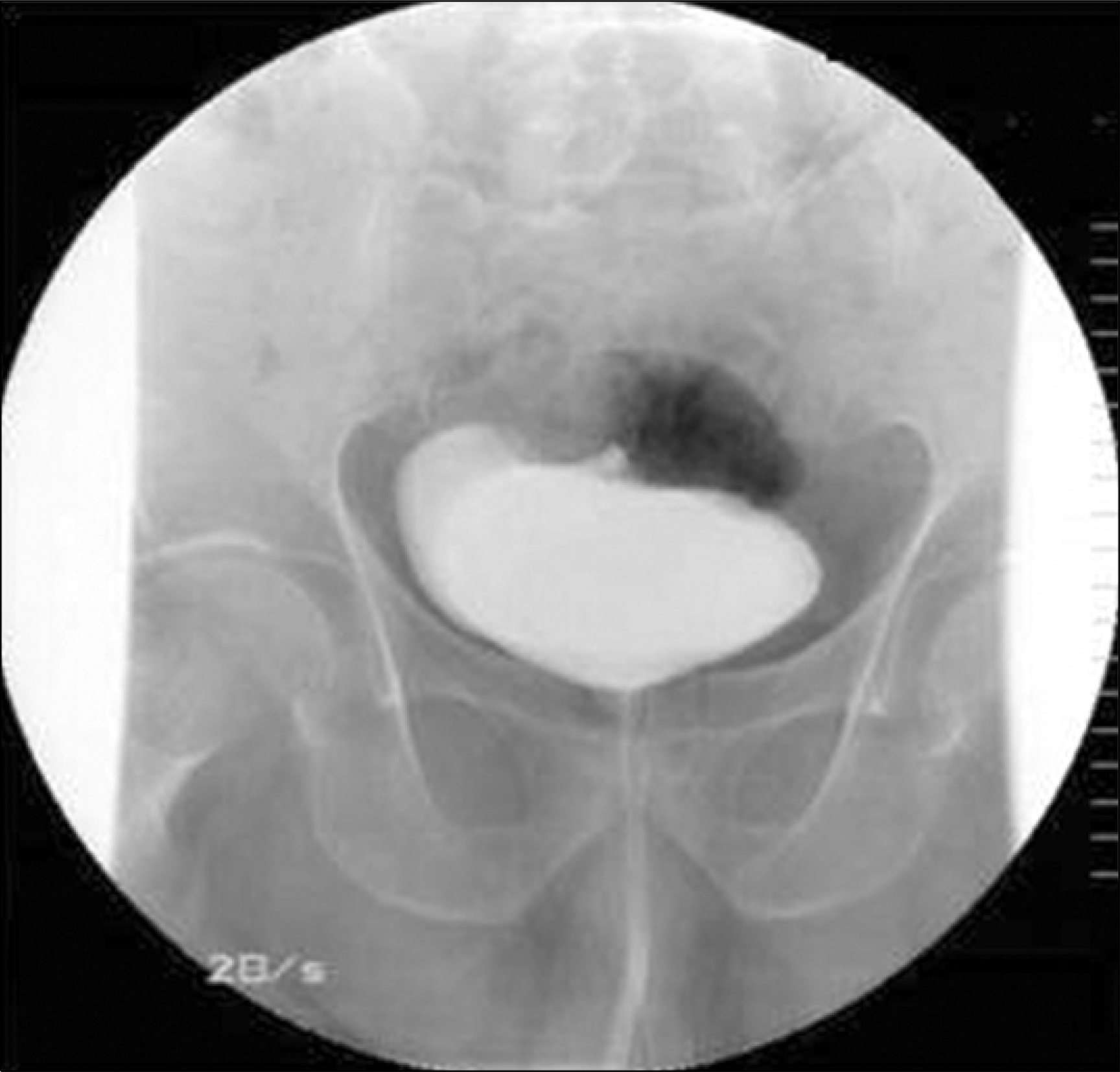 | Fig. 1.Cystogram finding. About 300 cc of contrast media was filled in the urinary bladder and no contrast leakage was seen. The margin of the urinary bladder was relatively regular. |
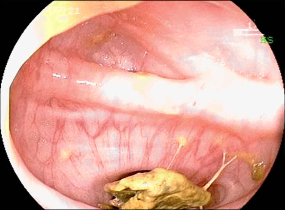 | Fig. 2.Sigmoidoscopic finding. At the sigmoid colon-25 cm from anal verge, a large diverticulum with impacted stool was noted. |
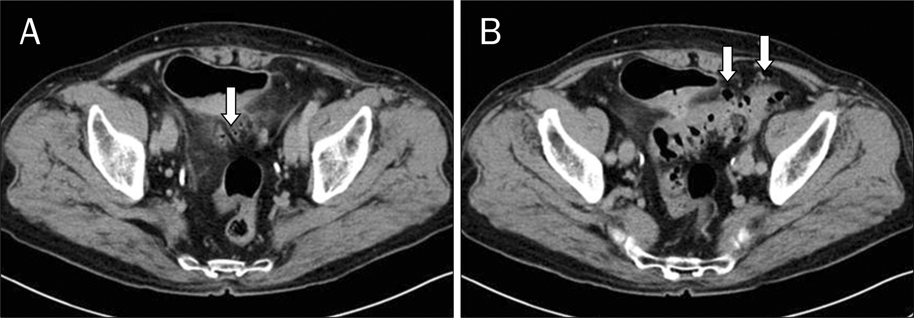 | Fig. 3.CT scan finding. (A) There was about 1.5 cm sized fistular tract between the sigmoid colon and posterior wall of the urinary bladder (white arrow). Perilesional bladder wall thickening and fat infiltrations were also noted. (B) Multiple diverticula were seen in the sigmoid colon (white arrows). |
 XML Download